
GITNUXSOFTWARE ADVICE
Healthcare MedicineTop 10 Best Medical 3D Software of 2026
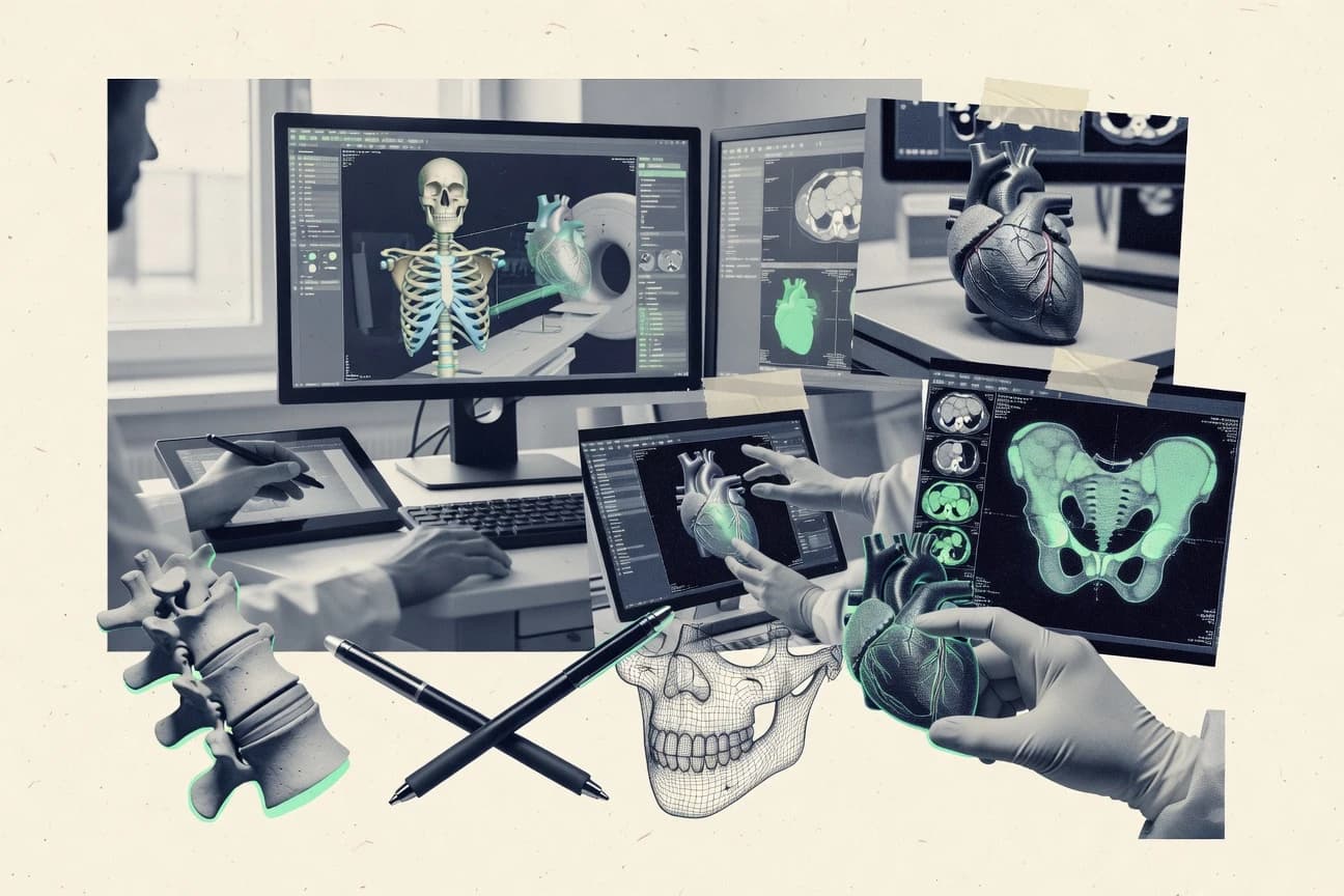
Top 10 medical 3d software ranked for imaging, segmentation, and planning, with editorial comparisons of tools like 3D Slicer and Mimics.

How we ranked these tools
Core product claims cross-referenced against official documentation, changelogs, and independent technical reviews.
Analyzed video reviews and hundreds of written evaluations to capture real-world user experiences with each tool.
AI persona simulations modeled how different user types would experience each tool across common use cases and workflows.
Final rankings reviewed and approved by our editorial team with authority to override AI-generated scores based on domain expertise.
Score: Features 40% · Ease 30% · Value 30%
Gitnux may earn a commission through links on this page — this does not influence rankings. Editorial policy
Horos is the most practical pick for DICOM imaging teams who want local segmentation plus reliable 3D mesh export for planning, while 3D Slicer is the better fit for teams needing interactive segmentation with automated, scriptable export for research.
Editor’s top 3 picks
Three quick recommendations before you dive into the full comparison below — each one leads on a different dimension.
Horos
Voxel-based segmentation with HU thresholding tailored to CT workflows inside a DICOM-first viewer.
Built for fits when DICOM imaging teams need local segmentation and mesh export for planning..
3D Slicer
Editor pickScriptable pipeline control via Python lets the same segmentation workflow run across studies with consistent parameters.
Built for fits when teams need interactive segmentation plus automated, scriptable export for planning and research..
Materialise Mimics
Editor pickLandmark-based registration plus measurement stays within the segmentation authoring workflow for planning-grade alignment.
Built for fits when clinical teams need repeatable image-to-geometry planning models across many cases..
Comparison Table
Horos
SMBOpen-source medical image viewer for macOS with 3D capabilities.
Voxel-based segmentation with HU thresholding tailored to CT workflows inside a DICOM-first viewer.
Horos is used for DICOM review, multi-planar reformation, and task-focused annotation that supports clinical image interpretation before 3D export. Segmentation can be driven by HU thresholding for CT volumes and then refined into surface representations for review and measurement. For 3D work, the reconstruction workflow commonly culminates in mesh output for external planning steps.
A notable tradeoff is that Horos is desktop-centric and does not provide a built-in, centralized server workflow for multi-site automation. It fits clinics that already manage imaging inputs as DICOM and want local control over segmentation and export for surgical planning output.
- +Strong DICOM workflow with multi-planar viewing and annotation tools
- +Voxel-based segmentation supports thresholding and region growth refinement
- +3D export output includes mesh formats suitable for downstream planning
- +Plugin extensibility supports niche imaging and processing workflows
- –Desktop-first model limits centralized orchestration across multiple sites
- –Advanced 3D segmentation workflows require time to master
- –Automation depends heavily on add-ons rather than built-in pipelines
- –High-fidelity mesh workflows may require external tools for cleanup
Radiology researchers
CT segmentation for anatomy measurements
Consistent region quantification
Surgical planning teams
Patient-specific 3D planning models
Faster planning iterations
Show 2 more scenarios
Biomedical engineers
Mesh prep for simulations
Reduced manual rework
Export segmentation-derived meshes into external pipelines for processing and simulation-ready geometry.
Imaging operations groups
Standardized local export pipeline
More uniform outputs
Use a repeatable DICOM view and segmentation process to generate consistent STL deliverables.
Best for: Fits when DICOM imaging teams need local segmentation and mesh export for planning.
3D Slicer
vertical specialistOpen-source platform for medical image informatics and 3D visualization.
Scriptable pipeline control via Python lets the same segmentation workflow run across studies with consistent parameters.
3D Slicer provides a single UI for segmentation and planning tasks, with a modular architecture that exposes functionality as installable extensions. Common workflows include manual and semi-automatic segmentation, surface reconstruction, and multi-planar reformation for cross-checking anatomy. It can bring DICOM data through its import stack and work with DICOM RT structures for lesion labeling and anatomical outlines. Python automation supports parameterized routines for loading data, running segmentation steps, and exporting outputs.
A clear tradeoff is governance overhead when workflows depend on extra extensions and custom scripts, since consistent installs across workstations require coordination. 3D Slicer fits teams that need interactive segmentation for surgical planning cases while also running batch jobs for research cohorts. It is also a practical fit when clinicians and researchers need to inspect results in multiple views and then export meshes for downstream use.
- +Python scripting enables reproducible batch segmentation and export workflows
- +Extension ecosystem adds specialized tools without changing the core app
- +Integrated registration and multi-view inspection support iterative planning
- +Active segmentation toolset covers voxel editing and region labeling
- –Extension and script dependencies increase configuration and validation burden
- –Complex pipelines can require workflow knowledge and repeatable conventions
Radiology research teams
Batch lesion segmentation for cohorts
Consistent labels across datasets
Surgical planning clinicians
Patient-specific anatomy alignment and review
Fewer manual review loops
Show 1 more scenario
Imaging IT administrators
Controlled workstation deployment for projects
Reduced variability between users
Standardize extension sets and scripted workflows across a team of imaging users.
Best for: Fits when teams need interactive segmentation plus automated, scriptable export for planning and research.
Materialise Mimics
enterpriseSoftware for creating 3D models from medical image data.
Landmark-based registration plus measurement stays within the segmentation authoring workflow for planning-grade alignment.
Materialise Mimics is used to create patient-specific 3D models from CT and MR studies using voxel-based segmentation, thresholding, and editing in axial, coronal, and sagittal views. It then supports surface extraction for mesh outputs that feed surgical planning, engineering review, and fabrication pipelines. The practical differentiator is the end-to-end focus from image segmentation to production-ready geometry, with strong tooling for measurement, labeling, and alignment tasks inside the same authoring environment.
A tradeoff is that Mimics can require more time to set up a repeatable segmentation workflow than lighter editors, especially when teams need consistent parameterization across organs or scanners. Mimics fits settings where the same segmentation logic must be applied across multiple cases, such as orthopedic preoperative planning that depends on stable landmark registration and consistent export geometry for downstream use.
- +Strong DICOM-driven segmentation workflow with multi-planar editing
- +Tight measurement and annotation support for planning-ready models
- +Better control of surface extraction and mesh cleanup than basic viewers
- +Repeatable project workflows suited to batch-like clinical case production
- –Requires segmentation methodology discipline to keep results consistent
- –Advanced mesh prep often depends on additional Materialise tools
Orthopedic planning teams
Femur and pelvis models from CT
Consistent planning geometry export
Maxillofacial surgeons
Craniofacial segmentation for surgical guides
Production-ready guide models
Show 1 more scenario
Clinical imaging analysts
Standardized segmentation across scanner types
More consistent segmentation outputs
Uses repeatable segmentation steps to reduce variability between studies during model creation.
Best for: Fits when clinical teams need repeatable image-to-geometry planning models across many cases.
Brainlab
enterpriseSoftware for digital surgery and 3D surgical planning.
Intraoperative navigation alignment workflows built to carry plan-to-procedure context with controlled registration steps.
Brainlab combines medical imaging, segmentation tools, and surgical planning workflows into a single environment for patient-specific 3D visualization. Its suite supports DICOM-based exchange and interop workflows used across radiology and image-guided therapy.
Brainlab also includes 3D model generation and registration tools that feed intraoperative navigation use cases. It is positioned for teams that need repeatable planning steps with controlled configuration across cases.
- +Strong surgical planning workflow designed for image-guided navigation handoff
- +DICOM-focused integration supports structured clinical exchange pathways
- +3D segmentation and labeling tooling geared to clinical case reuse
- +Registration tools support alignment for longitudinal and intraoperative contexts
- –Complex workflow setup can slow down teams without dedicated admin support
- –Advanced planning steps often require training beyond basic imaging tasks
- –Segmentation flexibility depends on configured tools for each anatomy workflow
- –Project organization can feel heavyweight compared with lightweight editors
Best for: Fits when imaging, segmentation, and surgical planning must stay consistent from DICOM import to navigation-ready models.
Visage Imaging
enterpriseEnterprise imaging platform with 3D advanced visualization.
Workflow-guided segmentation and measurements for repeatable surgical planning case review.
Visage Imaging performs medical 3D visualization, segmentation, and surgical planning workflows from clinical imaging inputs. It is used to generate patient-specific 3D models and measurement outputs with tightly guided tools for anatomy labeling and structured review.
The workflow center is around handling clinical image series, creating derived 3D representations, and exporting results to downstream documentation or fabrication pipelines. Integration depth shows most clearly in how the product fits into imaging-centric environments and in how repeated tasks can be standardized through configuration and automation.
- +Configurable clinical workflows for consistent segmentation and measurement steps
- +Strong support for medical 3D visualization suited to review and planning
- +Designed around repeatable cases with predictable derived outputs
- +Works well with imaging-centric operational requirements for study-to-result flow
- –Project setup can require governance discipline for consistent labeling outcomes
- –Specialized planning workflows may lag behind niche open toolkits
- –Automation depends on how workflows are packaged for each use case
- –Export and downstream formatting can be constrained by workflow boundaries
Best for: Fits when clinical teams need standardized 3D review and planning workflows across many studies.
Mirada Medical
vertical specialistSoftware for medical image analysis and fusion.
Workflow configuration for study-scale automation that ties segmentation results to review outputs and downstream documentation artifacts.
Mirada Medical focuses on medical 3D image analysis workflows that combine segmentation, measurements, and study-level reporting for clinical and research use. It is distinct in how it frames imaging tasks around repeatable pipelines that can be configured for different modalities and anatomy.
Core capabilities center on segmentation support with downstream review artifacts and export paths for 3D visualization and clinical documentation. For teams with integration needs, Mirada Medical typically fits best when DICOM-based imaging inputs and automated processing steps must be coordinated across studies.
- +Configurable processing pipelines for repeatable segmentation and measurement workflows
- +Strong support for review artifacts that help verify segmentation results
- +Designed for clinical study contexts with output geared toward reporting and downstream use
- +Integration-oriented workflow structure for coordinating imaging tasks across studies
- –Segmentation accuracy depends heavily on the provided models and configuration
- –Advanced customization can require specialist workflow configuration rather than simple point-and-click edits
- –Export options vary by workflow, which can complicate standardized mesh pipelines
- –Complex multi-step automation can be harder to debug than simpler single-tool editors
Best for: Fits when imaging teams need repeatable segmentation and measurement workflows with controlled outputs for study operations.
Surgical Theater
vertical specialistVR and 3D software for surgical planning and rehearsal.
Clinic-oriented surgical planning workspace that couples anatomy labeling and manual refinement with scene verification steps.
Surgical Theater focuses on end-to-end patient-specific 3D modeling for surgical planning, with a workflow designed around anatomy-focused scene building rather than generic mesh tools. The software supports DICOM ingestion, segmentations, and exports into common 3D assets for downstream planning and visualization.
Its planning workspace emphasizes manual refinement tools and spatial verification steps that fit clinic review sessions. Compared with tools like 3D Slicer and Mimics, Surgical Theater typically maps more directly to a surgical workflow, with fewer general-purpose reconstruction paths.
- +Surgical planning workflow is organized around patient-specific scene review
- +DICOM import supports clinical imaging handoffs into a planning workspace
- +Segmentation refinement tools support practical operator corrections
- +Export formats support downstream visualization and asset-based planning
- –Less flexible than 3D Slicer for custom segmentation algorithm prototyping
- –API and automation surface are not as clearly aligned to large-scale pipelines
- –Mesh processing depth is narrower than dedicated model editing toolchains
- –Workflow benefits depend on consistent input quality and acquisition protocols
Best for: Fits when clinical teams need fast patient-specific 3D planning from DICOM, with operator-guided refinement and export for review.
Anatomage
vertical specialist3D anatomy visualization software and virtual dissection tables.
Anatomage’s integrated anatomical landmarking workflow drives measurements and patient-specific 3D scene updates in one interface.
Anatomage delivers medical 3D visualization and modeling tools for anatomical inspection, measurements, and surgical planning workflows. Its core strength is patient-specific anatomical modeling with interactive landmarking, segmentation refinement, and downstream export for 3D workflows.
The application also supports common medical imaging handling such as DICOM-based data and multi-planar views that feed a single 3D scene. Compared with toolchains like 3D Slicer or Mimics, Anatomage focuses on a tightly integrated workstation workflow instead of a plugin-heavy build-from-scratch pipeline.
- +Integrated 3D inspection with measurement tools tied to landmark workflows
- +Patient-specific modeling workflow that stays within one interactive scene
- +DICOM-oriented imaging workflow with multi-planar support and 3D synchronization
- +Export options designed for downstream surgical planning and 3D fabrication pipelines
- –API and automation surface are limited compared with extensible medical toolchains
- –Advanced automation for large batch segmentation is not its primary strength
- –Depth of governance controls for enterprise deployment is less transparent than dedicated platforms
- –Some segmentation workflows depend on manual refinement rather than fully scripted steps
Best for: Fits when clinical teams need guided 3D planning and measurement with minimal pipeline assembly.
Able Software 3D-DOCTOR
SMBSoftware for creating 3D models from CT MRI and ultrasound scans.
Interactive, case-focused 3D modeling workflow designed for iterative anatomical review and export-driven planning.
Able Software 3D-DOCTOR is a medical 3D viewer and modeling workflow for turning medical images into patient-specific 3D geometry for review and planning. It supports interactive segmentation workflows and exports common geometry outputs for downstream tools.
The workflow emphasizes repeatable planning steps on workstation scale rather than fully automated pipeline orchestration. Integration depth depends on how local imaging formats and exports are used to connect to PACS and external planning or printing steps.
- +Interactive segmentation and labeling workflows designed for clinical workstation use
- +Exports patient geometry for downstream planning and 3D printing pipelines
- +Handles multi-step 3D review workflows without forcing a scripting layer
- +Focused toolchain reduces configuration churn during planning sessions
- –Limited automation depth for end-to-end batch processing across large case loads
- –External integration relies heavily on import and export steps rather than native orchestration
- –Governance controls for multi-user deployments are not clearly positioned for enterprise RBAC needs
- –Mesh repair and simulation prep are not as specialized as dedicated planning ecosystems
Best for: Fits when radiology teams need workstation-based segmentation and repeatable 3D review outputs.
ITK-SNAP
vertical specialistOpen-source tool for 3D image segmentation and navigation.
Interactive region-growing and split editing tools designed for refining segmentation boundaries across views.
ITK-SNAP is a medical 3D imaging and segmentation tool built around fast interactive label creation and slice-by-slice review. It supports voxel-based workflows for segmenting structures from volumetric data and exporting results for downstream 3D analysis.
The workflow emphasizes anatomy-first navigation, including multi-planar views and segmentation propagation aids, rather than building a full surgical planning stack. Compared with generalist viewers, it focuses on annotation quality, reproducible segmentation passes, and interoperability through common medical imaging exports.
- +Voxel-based segmentation tools that support accurate boundary editing
- +Multi-planar review helps catch labeling errors across orthogonal slices
- +Segmentation workflow is optimized for fast iteration and rework
- +Export support fits common pipelines that expect standard geometry outputs
- –Limited built-in DICOM RT structure set and PACS orchestration compared with hospital stacks
- –No integrated surgical planning or simulation toolchain for end-to-end workflow
Best for: Fits when teams need fast, repeatable medical image segmentation and geometry export for analysis or planning.
Conclusion
After evaluating 10 healthcare medicine, Horos stands out as our overall top pick — it scored highest across our combined criteria of features, ease of use, and value, which is why it sits at #1 in the rankings above.
Use the comparison table and detailed reviews above to validate the fit against your own requirements before committing to a tool.
How to Choose the Right medical 3d software
Medical 3D software turns DICOM imaging into patient-specific geometry for segmentation and planning workflows, with the strongest emphasis on voxel-based CT handling, repeatable labeling, and geometry export. This buyer’s guide covers Horos, 3D Slicer, Materialise Mimics, Brainlab, Visage Imaging, Mirada Medical, Surgical Theater, Anatomage, Able Software 3D-DOCTOR, and ITK-SNAP.
The tool set is split between DICOM-first desktop workstations like Horos and ITK-SNAP, and larger clinical workflow platforms like Brainlab and Visage Imaging. The guide also includes scriptable, batch-oriented pipeline control in 3D Slicer and measurement plus landmark-aligned planning models in Materialise Mimics.
Medical 3D software for segmentation, measurement, and surgical planning from DICOM
Medical 3D software provides an authoring workflow for turning imaging volumes into segmentation masks and exportable 3D models used for surgical planning, review, and fabrication. Horos focuses on voxel-based segmentation with HU thresholding inside a DICOM-first viewer, with multi-planar viewing and annotation support for local refinement.
3D Slicer targets teams that need consistent results across studies using Python-driven pipeline control for reproducible batch segmentation and export. Across the broader set, differences show up in whether segmentation and planning steps stay inside one interactive workspace or require additional tooling and governance discipline to keep outcomes consistent across many cases.
Medical 3D software decision signals for segmentation, planning, and export
Segmentation quality determines whether planning measurements stay consistent, and voxel-based HU thresholding support in Horos is built for CT workflows inside a DICOM-first viewer. Multi-planar viewing and annotation tools matter because boundary edits must be validated across orthogonal slices during clinical review.
DICOM-first segmentation and multi-planar validation
Horos uses voxel-based segmentation with HU thresholding in a DICOM-first viewer for local refinement. 3D Slicer also supports interactive multi-planar segmentation while staying scriptable for repeatable export.
Scriptable batch pipelines for reproducible segmentation
3D Slicer enables Python scripting so the same segmentation workflow runs across studies with consistent parameters. Mirada Medical provides workflow configuration for study-scale automation that ties segmentation results to review outputs and downstream documentation artifacts.
Planning-grade alignment inside the authoring workflow
Materialise Mimics pairs landmark-based registration with measurement and keeps alignment inside segmentation authoring for planning-grade models. Brainlab focuses on intraoperative navigation alignment workflows that carry plan-to-procedure context through controlled registration steps.
Workflow-guided case review and measurement standardization
Visage Imaging offers configurable clinical workflows that guide segmentation and measurements for standardized surgical planning case review. Surgical Theater organizes the planning workspace around patient-specific scene review with anatomy labeling and manual refinement plus scene verification steps.
Inline landmarking and interactive patient-specific scene updates
Anatomage integrates anatomical landmarking so measurements and patient-specific 3D scene updates occur within one interactive interface. Able Software 3D-DOCTOR provides iterative anatomical review with export-driven planning built around interactive segmentation and labeling workflows.
Interactive boundary refinement tools with fast editing loops
ITK-SNAP delivers region-growing and split editing designed for refining segmentation boundaries across views. Horos complements CT segmentation with voxel-based HU thresholding and region growth refinement for boundary refinement loops.
How to choose medical 3D software by workflow control and automation depth
Start by classifying how the team needs to control segmentation variation across many studies. A DICOM-first workstation approach like Horos fits teams that want interactive voxel-based CT thresholding and local refinement. A script-driven approach like 3D Slicer fits teams that need the same segmentation logic executed consistently across studies.
Pick the control model for segmentation consistency
If consistent CT segmentation starts with local HU thresholding and refinement in a DICOM-first viewer, choose Horos for voxel-based thresholding plus region growth refinement. If segmentation consistency must be enforced via repeatable execution across studies, choose 3D Slicer because Python scripting supports reproducible batch segmentation and export workflows.
Decide whether landmark registration must stay inside segmentation
If teams need planning-ready alignment created as part of the same authoring workflow, choose Materialise Mimics because landmark-based registration and measurement stay within segmentation authoring. If navigation handoff is the central requirement, choose Brainlab because its intraoperative navigation alignment workflows keep plan-to-procedure context across controlled registration steps.
Match governance needs to workflow configurability
If the operational goal is standardized case review with guided segmentation and measurements, choose Visage Imaging because it provides configurable clinical workflows for repeatable surgical planning case review. If the goal is study-scale automation that ties segmentation to review artifacts and downstream documentation, choose Mirada Medical for workflow configuration that controls outputs.
Choose the authoring workspace philosophy for planning delivery
If planning delivery depends on an operator-guided patient-specific scene with labeling plus verification steps, choose Surgical Theater because scene review organizes manual refinement and export for review. If planning depends on interactive landmark-driven measurement and patient-specific scene updates with minimal pipeline assembly, choose Anatomage because the landmarking workflow drives the 3D scene updates.
Plan for where custom algorithms and extensions will come from
If custom segmentation logic must be implemented and maintained inside an extensible toolkit, choose 3D Slicer because its extension ecosystem adds specialized tools without changing the core app. If segmentation workflow prototyping is not a priority and the main task is iterative boundary refinement, choose ITK-SNAP because its split editing and region-growing tools focus on interactive boundary edits across views.
Assess whether advanced planning steps require dedicated training time
If the workflow includes complex planning steps that can slow down without admin support, treat Brainlab’s complex workflow setup as a factor and budget training time for controlled registration and navigation handoff. If the workflow includes advanced segmentation steps that take time to master, treat Horos’s advanced 3D segmentation workflows as a learning curve that needs process time.
Who should buy medical 3D software for segmentation and surgical planning work
DICOM imaging teams need predictable segmentation outputs that can feed planning models and exports, and Horos is built around voxel-based HU thresholding inside a DICOM-first viewer. Research and engineering teams also need reproducible workflows across studies, and 3D Slicer’s Python-driven pipeline control supports that consistency.
DICOM imaging teams doing local CT segmentation
Horos fits teams that need voxel-based segmentation with HU thresholding plus multi-planar viewing and annotation for local refinement before exporting planning geometry.
Research teams running repeatable segmentation across studies
3D Slicer fits teams that require Python-scripted consistency because the same segmentation workflow can run across studies with reproducible batch export.
Clinical teams producing planning-ready alignment models
Materialise Mimics fits clinical workflows that require landmark-based registration and measurement staying inside the segmentation authoring process for planning-grade models.
Study operations teams standardizing outputs for review and documentation
Mirada Medical fits operational workflows that need study-scale automation because it ties segmentation results to review outputs and downstream documentation artifacts.
Navigation and intraoperative alignment teams
Brainlab fits teams that need image-guided navigation continuity because its intraoperative navigation alignment workflows carry plan-to-procedure context through controlled registration steps.
Common medical 3D software buying pitfalls that break planning consistency
A common failure mode is underestimating configuration discipline when a tool relies on workflow governance to keep outputs consistent. Visage Imaging requires governance discipline for consistent labeling outcomes, and 3D Slicer can add validation burden through extensions and scripts that must be kept aligned with conventions.
Choosing a workflow-guided platform without planning for labeling governance
Visage Imaging and Visage Imaging-style workflows require project setup governance discipline to keep labeling outcomes consistent across many studies. Budget process time for consistent configuration before scaling case review.
Assuming automation depth without validating extension and script dependencies
3D Slicer can increase configuration and validation burden because extensions and scripts are part of the pipeline. Treat the pipeline as a maintained artifact with tested conventions across studies.
Building an end-to-end planning workflow on a segmentation-only tool
ITK-SNAP provides interactive boundary editing but has limited built-in DICOM RT structure set and PACS orchestration compared with hospital stacks. Pair segmentation editing tools with a separate planning or orchestration workflow instead of expecting one integrated pipeline.
Selecting a desktop-first workflow when multi-site orchestration is required
Horos desktop-first design limits centralized orchestration across multiple sites. If distributed execution is required, select a platform designed for study-scale automation or pipeline control.
Overlooking how planning alignment steps affect handoff to navigation
Materialise Mimics keeps landmark registration and measurement inside segmentation authoring, while Brainlab focuses on navigation-ready registration handoff. Choose based on whether alignment must support intraoperative navigation context rather than only planning-grade measurement.
How We Selected and Ranked These Tools
We evaluated Horos, 3D Slicer, Materialise Mimics, Brainlab, Visage Imaging, Mirada Medical, Surgical Theater, Anatomage, Able Software 3D-DOCTOR, and ITK-SNAP using features at 40% weight and ease and value at 30% weight each. Features emphasized voxel-based CT handling, workflow organization for segmentation and planning, and export-oriented authoring support.
Ease emphasized how quickly teams can execute segmentation, measurement, and planning tasks without adding excessive setup burden. Horos earned the top rank because voxel-based segmentation with HU thresholding is tailored to CT workflows inside a DICOM-first viewer with multi-planar viewing and annotation tools for local refinement.
Frequently Asked Questions About medical 3d software
How do Horos, 3D Slicer, and Mimics handle DICOM-based segmentation into 3D geometry exports?
Which toolchain is better for repeatable automated segmentation pipelines across many studies: 3D Slicer or Mirada Medical?
When teams need intraoperative navigation alignment, where does Brainlab fit compared with Surgical Theater?
What breaks if segmentation must stay reproducible across reviewers when using ITK-SNAP versus Anatomage?
How do 3D Slicer and Horos differ in configuration depth for CT workflows using thresholding?
Which application supports RT structure handling alongside voxel-based segmentation: 3D Slicer or Mirada Medical?
How do Mirada Medical and Visage Imaging differ in study-level outputs after segmentation?
What security and deployment approach issues typically arise when comparing HIPAA-focused on-premise needs across Horos, 3D Slicer, and Brainlab?
How should teams choose between Surgical Theater and Anatomage when anatomy labeling must drive the rest of the workflow?
Tools reviewed
Primary sources checked during evaluation.
Referenced in the comparison table and product reviews above.
- Healthcare MedicineTop 10 Best Online Medical Software of 2026
- Healthcare MedicineTop 10 Best Medical Speech To Text Software of 2026
- Healthcare MedicineTop 10 Best Medical Device Inventory Tracking Software of 2026
- Healthcare MedicineTop 10 Best Medical Document Scanning Software of 2026
- Healthcare MedicineTop 10 Best Medical Equipment Planning Software of 2026
Keep exploring
Comparing two specific tools?
Software Alternatives
See head-to-head software comparisons with feature breakdowns, pricing, and our recommendation for each use case.
Explore software alternatives→In this category
Healthcare Medicine alternatives
See side-by-side comparisons of healthcare medicine tools and pick the right one for your stack.
Compare healthcare medicine tools→