GITNUXSOFTWARE ADVICE
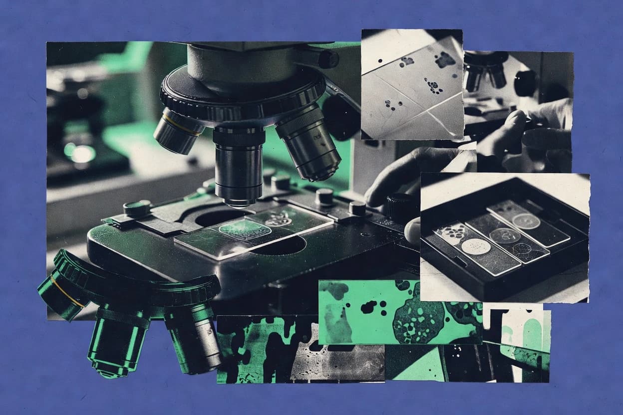
Science ResearchTop 10 Best Microscopy Software of 2026
Top 10 Microscopy Software ranked by imaging, analysis, and automation needs, with comparisons of VeraView, CellProfiler, and Napari.

How we ranked these tools
Core product claims cross-referenced against official documentation, changelogs, and independent technical reviews.
Analyzed video reviews and hundreds of written evaluations to capture real-world user experiences with each tool.
AI persona simulations modeled how different user types would experience each tool across common use cases and workflows.
Final rankings reviewed and approved by our editorial team with authority to override AI-generated scores based on domain expertise.
Score: Features 40% · Ease 30% · Value 30%
Gitnux may earn a commission through links on this page — this does not influence rankings. Editorial policy
Editor’s top 3 picks
Three quick recommendations before you dive into the full comparison below — each one leads on a different dimension.
VeraView
Provisioned API automation with RBAC and audit logs for specimen- and run-level governance.
Built for fits when mid-to-large imaging teams need governed metadata automation across microscopes..
CellProfiler
Editor pickObject classification and measurement export tied to segmentation outputs and object classes.
Built for fits when microscopy teams need repeatable workflow automation with exportable measurement schemas..
Napari
Editor pickLayer-based data model with a Python API that keeps images and label masks synchronized during editing.
Built for fits when teams need programmable visualization and iterative QC for multidimensional microscopy workflows..
Related reading
Comparison Table
This comparison table evaluates microscopy software across integration depth, focusing on how each tool connects with image formats, downstream pipelines, and existing analysis stacks. It also contrasts each tool’s data model and schema strategy, plus automation, API surface, and extensibility through scripts, plugins, and Python integration. Admin and governance controls are compared using provisioning support, RBAC patterns, and audit log coverage where available.
VeraView
image workflowsMicroscopy image management and analysis workflow software that supports automated organization, visualization, and quantification across image datasets.
Provisioned API automation with RBAC and audit logs for specimen- and run-level governance.
The core value comes from integration depth into imaging pipelines, where acquisition events, specimen identifiers, and analysis outputs remain linked inside a consistent schema. VeraView’s extensibility and automation surface supports workflow configuration for ingestion, validation, and handoff to analysis steps without breaking referential integrity. The data model is designed for repeated runs, so schema alignment matters when multiple microscopes and operators contribute data.
A tradeoff appears in the need to design the schema mappings and automation rules up front for each imaging program, because governance and auditability depend on consistent identifiers. VeraView fits when teams already have structured metadata at acquisition time and want controlled throughput into analysis systems. It also fits when cross-site access needs RBAC and audit logs tied to specimen-level records.
- +Schema-driven microscopy records preserve links from acquisition to analysis outputs
- +API-first automation supports pipeline orchestration and programmatic configuration
- +RBAC and audit log coverage supports controlled access to specimen and run history
- –Schema mapping work can be non-trivial for legacy metadata formats
- –Automation configuration requires clear naming and identifier conventions
Facility operations leads at multi-instrument microscopy centers
Standardize specimen tracking across shared microscopes and staff roles.
Operators and administrators reduce manual reconciliation and can audit specimen provenance during reviews.
Bioinformatics and imaging platform engineers
Connect acquisition, analysis, and downstream reporting using API-driven workflows.
Teams automate end-to-end metadata propagation without custom glue code that breaks linkage.
Show 2 more scenarios
Quality and compliance teams in regulated lab environments
Maintain traceability for specimen handling and analysis decisions.
Audits and internal reviews can verify who changed what and when at specimen and run granularity.
The governed data model supports audit logging and role-based access so changes to run and specimen records are traceable. Configuration controls help enforce consistent provenance fields across imaging sessions.
Research program managers coordinating cross-team imaging studies
Enable controlled sharing of imaging datasets across projects and groups.
Program managers can approve dataset readiness based on validated metadata instead of manual spot checks.
VeraView’s access controls and structured metadata support consistent retrieval by project, instrument, and specimen identifiers. Automation can create repeatable onboarding steps for new experiments while keeping the same schema.
Best for: Fits when mid-to-large imaging teams need governed metadata automation across microscopes.
CellProfiler
high-throughputOpen-source software for high-throughput microscopy image analysis that performs batch processing, feature extraction, and exportable results.
Object classification and measurement export tied to segmentation outputs and object classes.
Teams that need reproducible, multi-step microscopy analysis benefit from CellProfiler’s module pipeline and parameterized execution. Workflows can capture preprocessing, segmentation, feature extraction, and measurement export, with outputs organized by object class and image set identifiers. Integration depth is strongest for analysis orchestration through its scriptable interface and exported data tables rather than for governance-first enterprise systems.
A tradeoff appears when labs require strict RBAC, fine-grained audit logs, and centralized provisioning because CellProfiler workflows are typically managed by file-based projects and execution scripts. For example, a single lab or small microscopy core can standardize analysis runs across instruments by sharing the same pipeline configuration and exported schema. For cross-lab administration and access controls, external orchestration and dataset management often carry more of the governance load.
- +Workflow pipelines encode preprocessing, segmentation, and measurement steps in one run
- +Object-class measurement tables support structured downstream analytics
- +Scriptable execution supports batch throughput across image sets
- +Extensibility via custom modules enables domain-specific segmentation and features
- –RBAC and audit log controls are not built as centralized admin features
- –Workflow sharing often relies on files and execution environment alignment
- –Enterprise data governance integrations are limited compared with app-layer analytics stacks
Microscopy core facilities
Standardize analysis for recurring assays across multiple imaging runs and operators
Consistent per-image-set features that reduce operator-to-operator variation in QC decisions.
Bioinformatics and imaging platform engineers
Integrate segmentation and feature extraction into automated batch processing
Higher throughput with predictable inputs and schema-aligned outputs for downstream services.
Show 2 more scenarios
Drug discovery teams running phenotypic screens
Extract morphological and intensity features for hit triage at scale
Decision-ready feature sets that support reproducible hit selection and model training.
CellProfiler pipelines can segment nuclei and cells, compute features for each object class, and export a structured feature matrix. Feature extraction consistency across plates and batches improves the reliability of screening models and review.
Research groups building custom segmentation methods
Add domain-specific preprocessing or measurement logic for specialized microscopy modalities
Domain-specific features delivered through the same run orchestration and measurement schema.
Custom modules allow teams to extend the pipeline for specialized segmentation targets and measurements. The existing data model for object classes and measurements keeps custom outputs aligned with standard exports.
Best for: Fits when microscopy teams need repeatable workflow automation with exportable measurement schemas.
Napari
interactive viewerInteractive n-dimensional microscopy image viewer for rapid exploration and segmentation with Python plugin support.
Layer-based data model with a Python API that keeps images and label masks synchronized during editing.
The integration depth is driven by Napari’s layer abstraction and its Python API, which lets microscopy workflows share the same in-memory objects across visualization, segmentation, and measurement. Layers can represent images, labels, points, and shapes, so the viewer can mirror common microscopy preprocessing steps without forcing a proprietary file schema. Extensibility is achieved through a plugin architecture that adds both UI components and processing functions, which supports repeatable analysis workflows across projects.
A tradeoff is that governance and multi-user controls are not the primary focus since Napari runs as an interactive desktop application or within a notebook environment. Teams still gain control depth when they pair Napari with external orchestration and storage, because the viewer can act as the front end for deterministic analysis code. Napari fits most when throughput bottlenecks are visual QC and iterative annotation on large 2D or 3D datasets, not when centralized admin and RBAC are required inside the viewer.
- +Python API drives automation and lets plugins modify the live layer model
- +Consistent layer types unify images, labels, points, and shapes for microscopy workflows
- +Plugin ecosystem adds segmentation, tracking, and tooling without rewriting the viewer
- +Notebook and script integration supports reproducible interactive QC sessions
- –No built-in RBAC or audit log for multi-user governance inside the app
- –Desktop interactivity can limit centralized administration for large labs
- –Automation relies on external pipeline orchestration for job control
Microscopy image analysis teams writing Python-based pipelines
Run segmentation experiments and visualize label edits while keeping algorithm code in the same runtime
Faster experiment iteration with fewer ad hoc exports and consistent label handling across steps.
Core facility scientists coordinating QC across multiple projects
Standardize interactive QC checks for 2D and 3D microscopy batches using reusable scripts and plugins
More consistent QC decisions through a repeatable configuration and processing entry point.
Show 2 more scenarios
Lab engineers building extensible analysis tools for internal use
Add custom measurement, atlas navigation, or data import into a microscopy workflow using the plugin surface
Controlled extensibility where internal tools stay coupled to the viewer’s data model and configuration.
Custom plugins can register new layer behaviors and UI elements, and they can operate directly on the layer model. This supports extensibility without changing core viewer code, which helps teams version their microscopy tooling around their own schema.
R&D teams integrating visualization into automated pipelines
Use Napari as a view layer in a headless processing workflow that generates derived images and labels
Improved throughput by reducing manual file handling and by aligning visualization inputs with pipeline outputs.
External automation can create layers from pipeline outputs and render intermediate results for review, while the analysis remains in orchestration code. The plugin ecosystem can add specialized processing steps when intermediate visualization requires domain-specific operations.
Best for: Fits when teams need programmable visualization and iterative QC for multidimensional microscopy workflows.
Icy
bioimage platformOpen-source bioimage analysis platform for microscopy with plugins, interactive segmentation, and batch processing support.
Icy plugin framework for adding new image processing pipelines and reusing them across sessions
Icy targets microscopy image analysis with a plugin-driven architecture that exposes analysis workflows as reusable components. It stores imaging data and derived results through a viewer-centric data model rather than a strict external schema, which affects how systems integrate downstream.
Automation is achieved via scripting hooks and batch execution patterns, with an extensibility surface that supports custom processing modules. Integration depth depends on how analysis outputs can be serialized into interoperable formats and fed into lab pipelines using its extension points.
- +Plugin architecture supports custom microscopy analysis modules without replacing the core
- +Scripting and batch processing enable unattended throughput runs over image sets
- +Viewer-oriented data handling simplifies interactive quality control before export
- –Data model is viewer-centric, which limits strict external schema alignment
- –Automation coverage is extension-dependent, so API surface varies by workflow type
- –Governance controls like RBAC and audit logging are not documented as first-class features
Best for: Fits when teams need extensible, workflow automation for microscopy analysis with light integration.
Python Scientific Stack (NumPy, SciPy, scikit-image)
library toolkitCommon microscopy image-processing toolchain implemented in Python for segmentation, filtering, morphology, and feature extraction with programmatic control.
scikit-image offers scikit-image.measure and skimage.registration APIs for segmentation metrics and alignment.
Python Scientific Stack provides array-first imaging computation using NumPy, signal and optimization routines via SciPy, and segmentation, registration, and morphology tools through scikit-image. In microscopy workflows, it fits as an integration layer where analysis code and image pipelines share the same in-memory data model.
The automation surface is the Python API, which enables custom batch processing, reproducible transforms, and extensibility through user-defined functions and plugins. Admin and governance controls are limited to what can be built around Python execution, since scikit-image and its dependencies do not ship built-in RBAC or audit logging.
- +NumPy arrays unify image, masks, and metrics in one data model
- +Consistent Python API supports scripted pipelines and reproducible transforms
- +scikit-image covers segmentation, registration, morphology, and feature extraction
- +SciPy adds optimization, filtering, and signal tools for upstream preprocessing
- +Extensibility through user code lets pipelines run inside existing lab systems
- –No native RBAC, audit logs, or admin governance for shared environments
- –Workflow orchestration requires external tooling for scheduling and retries
- –Large datasets can stress RAM since core operations keep data in memory
- –Version drift across the scientific stack can break long-running pipelines
Best for: Fits when microscopy teams need Python-controlled analysis throughput with custom automation and minimal platform constraints.
ImageJ
plugin-based analysisExtensible microscopy image analysis platform with a large plugin ecosystem for measurement, segmentation, and visualization.
ImageJ macro language and Java plugin API for automation and custom analysis steps.
ImageJ is a microscopy analysis tool with deep plugin integration and a scriptable workflow via ImageJ macro language. Its data handling centers on image and ROI objects plus a persistent image processing pipeline, which supports repeatable measurement across batches.
Automation relies on macro execution, command-line batch processing, and plugin APIs rather than a separate orchestration service. Extensibility is driven by Java plugins and macro scripting, but governance controls like RBAC and audit logs are not built into the core desktop model.
- +Plugin API supports custom microscopy analysis modules in Java
- +Macro language enables repeatable batch processing for high throughput
- +Command-line batch execution supports headless pipelines and scripting
- +ROI and measurement tools standardize quantitative outputs across images
- –Core app model lacks built-in RBAC and centralized governance controls
- –Automation surface centers on macros and plugins rather than HTTP APIs
- –Large pipelines often require manual orchestration across plugins and scripts
- –Data model is tied to image objects, limiting schema-driven data governance
Best for: Fits when lab teams need scriptable image analysis with plugin extensibility, not enterprise governance.
OmeTiff
file format toolingTooling for working with OME-TIFF microscopy files using structured metadata and compatibility checks across viewers and analysis code.
Ome-TIFF channel and metadata mapping that preserves imaging context through exports.
OmeTiff focuses on microscopy-ready TIFF handling and metadata preservation for downstream analysis pipelines. It centers a structured data model for images and channels so exports map cleanly into analysis tools.
The integration story relies on filesystem workflows and extensibility hooks rather than a broad enterprise API surface. Automation is possible through scripted conversion and batch processing patterns built around those data and metadata assumptions.
- +Keeps Ome-TIFF metadata aligned with channels and imaging parameters.
- +Batch-friendly TIFF conversion workflow supports higher throughput on datasets.
- +Extensibility supports custom scripts around the image and metadata schema.
- +Designed around microscopy data structures rather than generic image blobs.
- –Limited admin governance features like RBAC and audit logging.
- –Automation surface is more file-driven than API-driven.
- –Schema rigidity can complicate nonstandard acquisition metadata.
- –Integration with orchestration systems needs external glue code.
Best for: Fits when teams need consistent OME-TIFF metadata exports with scriptable batch throughput.
BigDataViewer (BDV)
large-volume viewerMicroscopy visualization software for large multidimensional datasets using tiled rendering and interactive navigation.
Multiresolution tiled pyramids for interactive big-image navigation
BigDataViewer focuses on large microscopy image viewing inside ImageJ-based workflows, with multiscale navigation over tiled data. Its integration depth comes from native hooks to ImageJ ecosystems and support for common microscopy formats and coordinate-driven browsing.
The data model centers on tiled, spatially indexed pyramids that support interactive throughput for big datasets. Automation and API surface are limited compared with server-grade viewers, so extensibility depends more on ImageJ plugin patterns and batch processing than on provisioning or programmatic governance.
- +Built for ImageJ workflows and plugin interoperability
- +Tiled multiresolution pyramid model improves large-image navigation
- +Coordinate-based browsing supports consistent spatial context
- +Batch-ready processing via ImageJ scripting patterns
- –Limited server-side automation and API depth
- –Weak RBAC and audit log controls for team governance
- –Provisioning and schema management require ImageJ-side conventions
- –Throughput is optimized for viewing more than programmatic pipelines
Best for: Fits when ImageJ-centered teams need fast multiscale microscopy visualization with minimal infrastructure.
StarDist
deep segmentationTrainable instance segmentation approach packaged as Python tooling for nuclei and cell boundary detection on microscopy images.
Star-convex instance segmentation model that predicts polygon parameters per object.
StarDist runs instance segmentation for microscopy images by converting nuclei or objects into star-convex polygons. The project provides model training and inference workflows driven by a clear image-to-label data model.
Integration depth is strongest inside the Python stack through scikit-image style I/O and a model-centric API surface for batch prediction. Automation and governance controls are limited because the repository centers on research code rather than multi-user administration features like RBAC or audit logs.
- +Instance segmentation output uses star-convex geometry suitable for microscopy objects
- +Python inference code fits analysis pipelines with array-based inputs and outputs
- +Model training workflow supports custom datasets and object classes
- –Automation surface is mostly code-run, not a managed service API
- –Multi-user governance features like RBAC and audit logs are not provided
- –Throughput tuning depends on the user’s pipeline code, not built-in job control
Best for: Fits when teams automate Star-convex instance segmentation in Python workflows with custom model training.
Ilastik
ML segmentationGraphical machine-learning image segmentation toolkit for learning pixel or object classifiers from microscopy training examples.
Interactive pixel classification training from annotations with feature computation and saved project state.
Ilastik fits microscopy groups that need interactive segmentation workflows where labels and features can be regenerated across experiments. It centers a data model made for image-dependent feature computation and supervised training from user annotations.
Automation is mainly achieved by reproducible project files that can be reloaded for batch processing and consistent model application. Integration depth is constrained by limited external API surface, so extensibility typically happens through workflow handoffs rather than programmatic pipeline control.
- +Supervised training from user labels using a structured feature pipeline.
- +Project files preserve workflows for repeatable batch segmentation runs.
- +Interactive feedback shortens the annotation to model iteration loop.
- +Exported predictions support throughput across many images.
- –Automation relies on project-driven execution rather than exposed APIs.
- –Programmatic schema control and data validation are limited for governed pipelines.
- –RBAC and audit log controls are not designed for centralized administration.
- –Integrating custom model logic requires manual workflow wiring.
Best for: Fits when microscopy labs need consistent supervised segmentation without deep pipeline governance or external APIs.
How to Choose the Right Microscopy Software
This buyer's guide covers microscopy software used for image management, visualization, analysis automation, segmentation, and metadata-driven workflows. It maps evaluation criteria to tools including VeraView, CellProfiler, Napari, Icy, ImageJ, OmeTiff, BigDataViewer, StarDist, Ilastik, and the Python Scientific Stack.
The guide focuses on integration depth, data model design, automation and API surface, and admin and governance controls. Each section points to concrete mechanisms in specific tools, including RBAC and audit logs in VeraView and layer-based editing with a Python API in Napari.
Microscopy workflow software that turns images and metadata into governed analysis records
Microscopy software coordinates microscopy image handling, segmentation or analysis steps, and the data outputs that later code or lab staff consume. Some tools center on exportable measurement schemas like CellProfiler, while others center on interactive layer editing like Napari with a Python API.
These tools solve the problem of keeping acquisitions, labels, derived measurements, and run context consistent across experiments and teams. They are used by imaging teams that need repeatable throughput, researchers who iterate on segmentation and QC, and facilities that need auditability for specimen and run history such as VeraView.
Integration, schemas, and governance controls for microscopy data pipelines
Microscopy software selection hinges on whether the tool can preserve a consistent data model from acquisition to analysis output. VeraView uses schema-driven microscopy records for specimen- and run-level traceability, while tools like Icy and ImageJ use viewer-centric or image-and-ROI models that change how strict schema governance can work.
Integration depth and automation surfaces determine whether pipelines can be orchestrated by external job systems or controlled by internal APIs. Governance controls determine whether multi-user access and change history can be audited for specimen and run records in shared environments.
Schema-driven microscopy records with specimen and run traceability
VeraView ingests acquisition metadata and specimen context and produces structured, queryable records that preserve links from acquisition to analysis outputs. This schema-driven approach is paired with RBAC and audit log coverage so teams can govern specimen and run history rather than only store files.
Automation and API surface for provisioning and orchestration
VeraView provides a provisioned API automation surface that supports programmatic configuration and pipeline orchestration. CellProfiler supports automation via scriptable execution across image sets, while Napari shifts automation to a Python plugin ecosystem where code can modify the live layer model.
Data model alignment from segmentation or labels to measurement exports
CellProfiler ties object classes to measurement tables exported for downstream analytics, which keeps segmentation outputs connected to quantitative results. StarDist and the Python Scientific Stack fit when the workflow expects array-based inputs and structured label outputs from model inference or segmentation metrics.
Layer and label model coherence for interactive QC workflows
Napari uses a layer-based data model where images and label masks remain synchronized during editing, and the Python API drives automation around that live model. This mechanism matters when iterative QC is part of the microscopy workflow rather than a separate post-processing step.
Extensibility via plugins and module ecosystems
ImageJ and Icy extend microscopy analysis through plugins, with ImageJ offering macro language and a Java plugin API for repeatable batch processing. Icy uses a plugin framework for reusable analysis pipelines, which supports custom segmentation modules without replacing the core viewer.
Admin and governance controls that support controlled access and audit trails
VeraView includes RBAC and audit logging for specimen- and run-level governance, which supports multi-user administration. Other tools like Napari, ImageJ, and CellProfiler lack centralized RBAC and audit log controls as first-class admin features, so governance often needs external systems.
A controlled decision path from microscopy data model to automation and governance
Start with the data model that must survive the workflow, not the viewing or segmentation UI. VeraView fits when specimen and run records must remain queryable under a schema, while CellProfiler fits when measurement tables tied to object classes must export cleanly for analytics.
Next, validate automation and API surface against the job control needed in the lab environment. VeraView emphasizes an API-first automation path with provisioning, while Napari and the Python Scientific Stack emphasize Python-driven code and plugin execution without built-in centralized governance.
Match the data model to how results must be stored and queried
Select VeraView when microscopy outcomes must be stored as structured, queryable records that preserve links from acquisition to analysis outputs. Select CellProfiler when the workflow output must be object-class measurement tables tied to segmentation steps so downstream analytics can consume consistent exports.
Verify the automation surface can run inside the required pipeline controller
Choose VeraView when external automation needs programmatic provisioning and API-driven pipeline orchestration. Choose CellProfiler when batch throughput must be expressed as workflow pipelines with scriptable execution, or choose Napari when automation is expected through Python plugins that modify the live layer model.
Plan for governance requirements across multiple users and facilities
Choose VeraView when RBAC and audit logs must cover specimen and run history for controlled access. If selecting Napari, ImageJ, or the Python Scientific Stack, plan governance using external access controls because these tools do not ship built-in centralized RBAC and audit logging.
Align extensibility approach with how analysis logic will change
Select ImageJ when analysis logic changes frequently and the toolchain must support macro language plus a Java plugin API for custom steps. Select Icy when reusable microscopy analysis pipelines should be delivered as plugins and batch runs should reuse those modules.
Validate import and export compatibility for imaging formats and metadata
Choose OmeTiff when OME-TIFF channel and metadata mapping must preserve imaging context through exports and conversions. Choose BigDataViewer when the priority is interactive multiresolution tiled pyramids for large datasets inside ImageJ-centered workflows.
Which microscopy teams benefit from which tool patterns
Microscopy tool choice depends on whether the team needs governed metadata automation, exportable analysis measurements, programmable QC visualization, or interactive segmentation training. VeraView targets structured records and governance for imaging teams, while Napari targets iterative QC through a layer-based model and a Python API.
The audience-fit guidance below follows each tool’s best-for focus so tool selection aligns with operational workflows rather than UI preferences.
Mid-to-large imaging teams needing governed metadata automation across microscopes
VeraView fits because it produces schema-driven microscopy records from acquisition metadata and specimen context and pairs that model with RBAC and audit logging for specimen and run history.
Microscopy teams needing repeatable analysis automation with exportable measurement schemas
CellProfiler fits because workflow pipelines produce object-class measurement tables tied to segmentation outputs and support scriptable execution across image sets.
Teams needing programmable visualization and iterative QC for multidimensional microscopy workflows
Napari fits because its layer-based data model keeps images and label masks synchronized during editing and a Python API supports plugin-driven automation.
Microscopy teams needing extensible analysis pipelines with light integration
Icy fits because its plugin framework exposes reusable analysis pipelines and supports scripting and batch execution patterns without documenting centralized RBAC or audit logging.
Research groups automating segmentation or training with Python-centered pipelines
StarDist fits when star-convex instance segmentation inference and polygon parameters must be automated in Python, while Ilastik fits when supervised pixel classification training needs project-driven reproducible runs rather than an exposed external API.
Microscopy software pitfalls that break governance, automation, or schema consistency
Common selection failures come from assuming viewer-centric tools also provide governed schemas and admin controls. Napari, Icy, ImageJ, and the Python Scientific Stack emphasize interactive analysis and Python-driven computation, but they do not provide centralized RBAC and audit logging inside the core app model.
Other failures come from treating segmentation and measurements as disconnected outputs instead of a tied data model. Tools like CellProfiler connect object classes to measurement exports, while file-driven or model-centric tools require extra pipeline glue to keep schemas stable across steps.
Assuming built-in RBAC and audit logs exist in interactive analysis tools
VeraView provides RBAC and audit logging for specimen and run history, while Napari and ImageJ do not ship centralized admin governance controls. Governance-heavy environments should start with VeraView and only add external controls around tools that lack first-class RBAC and audit logs.
Treating exported results as optional rather than tied to a consistent data model
CellProfiler exports structured measurement tables tied to object classes, which keeps segmentation and quantification aligned across runs. Tools like Ilastik or StarDist can produce labels for automation, but the workflow must explicitly preserve the mapping from image inputs to predictions and measurement outputs.
Building pipelines around desktop-layer interactivity without planning job control
Napari is strong for a synchronized layer model and Python plugin automation, but job orchestration must come from external pipeline code because it does not provide server-grade automation control. For throughput automation, use CellProfiler workflow pipelines or a schema-first automation approach like VeraView.
Choosing file-driven format tooling without checking metadata governance needs
OmeTiff preserves OME-TIFF channel and metadata mapping through exports, which suits metadata-consistent microscopy file workflows. BigDataViewer supports tiled multiresolution viewing, but it provides limited API depth and weak governance controls, so it should not be treated as the system of record for specimen and run history.
How We Selected and Ranked These Tools
We evaluated each microscopy software option using features, ease of use, and value, and the overall rating is a weighted average where features carry the most weight. Ease of use and value each contribute the same share, which keeps automation capability and pipeline fit ahead of UI comfort.
VeraView ranks above lower-scored tools because it pairs schema-driven microscopy records with a provisioned API automation surface and RBAC plus audit logging for specimen and run-level governance. That combination lifts features and also improves practical ease of configuring controlled workflows across high-throughput imaging teams.
Frequently Asked Questions About Microscopy Software
How do Microscopy software tools handle metadata and data models for instrument and specimen context?
Which tools provide APIs or programmability for automation and provisioning of workflows?
How do microscopy tools integrate with existing analysis pipelines and storage systems?
What are the main tradeoffs between plugin-based desktop extensibility and enterprise governance controls?
Which tools support single sign-on and secure admin control of user permissions?
How should labs plan data migration when moving from desktop analysis to governed metadata pipelines?
Which software is best for multidimensional visualization and iterative QC on labeled data?
How do tools differ for batch throughput versus interactive exploration?
What common failure points appear when analysis outputs do not match a downstream expected data schema?
Conclusion
After evaluating 10 science research, VeraView stands out as our overall top pick — it scored highest across our combined criteria of features, ease of use, and value, which is why it sits at #1 in the rankings above.
Use the comparison table and detailed reviews above to validate the fit against your own requirements before committing to a tool.
Tools reviewed
Primary sources checked during evaluation.
Referenced in the comparison table and product reviews above.
Keep exploring
Comparing two specific tools?
Software Alternatives
See head-to-head software comparisons with feature breakdowns, pricing, and our recommendation for each use case.
Explore software alternatives→In this category
Science Research alternatives
See side-by-side comparisons of science research tools and pick the right one for your stack.
Compare science research tools→FOR SOFTWARE VENDORS
Not on this list? Let’s fix that.
Our best-of pages are how many teams discover and compare tools in this space. If you think your product belongs in this lineup, we’d like to hear from you—we’ll walk you through fit and what an editorial entry looks like.
Apply for a ListingWHAT THIS INCLUDES
Where buyers compare
Readers come to these pages to shortlist software—your product shows up in that moment, not in a random sidebar.
Editorial write-up
We describe your product in our own words and check the facts before anything goes live.
On-page brand presence
You appear in the roundup the same way as other tools we cover: name, positioning, and a clear next step for readers who want to learn more.
Kept up to date
We refresh lists on a regular rhythm so the category page stays useful as products and pricing change.