GITNUXSOFTWARE ADVICE
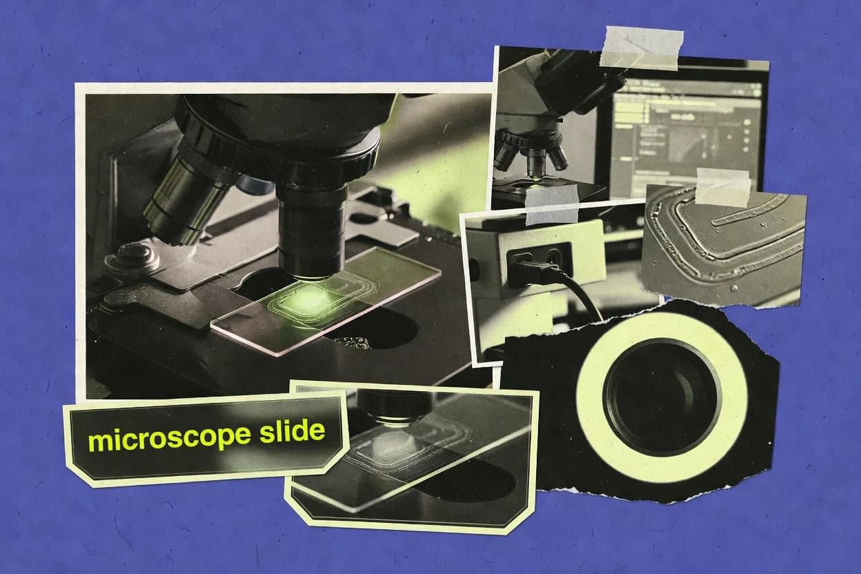
Science ResearchTop 10 Best Microscope Image Software of 2026
Top 10 ranking of Microscope Image Software for managing microscopy photos and analysis, with comparisons for labs and research workflows.

How we ranked these tools
Core product claims cross-referenced against official documentation, changelogs, and independent technical reviews.
Analyzed video reviews and hundreds of written evaluations to capture real-world user experiences with each tool.
AI persona simulations modeled how different user types would experience each tool across common use cases and workflows.
Final rankings reviewed and approved by our editorial team with authority to override AI-generated scores based on domain expertise.
Score: Features 40% · Ease 30% · Value 30%
Gitnux may earn a commission through links on this page — this does not influence rankings. Editorial policy
Editor’s top 3 picks
Three quick recommendations before you dive into the full comparison below — each one leads on a different dimension.
ilastik
Workflow-based training projects that link feature extraction, classifier training, and batch prediction.
Built for fits when microscopy teams need repeatable interactive segmentation with batch automation and controlled project configs..
CytoSMART
Editor pickAnnotation-first data model that preserves image metadata for repeatable microscopy review exports.
Built for fits when microscopy teams need governed image review workflows with automation-ready exports..
Omero alternatives: Terra Image
Editor pickMicroscopy dataset schema with metadata-driven organization and API automation hooks for processing pipelines.
Built for fits when labs need schema-governed microscopy automation with documented API integration and access control..
Related reading
Comparison Table
This comparison table evaluates microscope image software across integration depth, data model and schema design, and the automation and API surface for ingest, annotation, and analysis workflows. It also contrasts admin and governance controls such as RBAC, provisioning, and audit log coverage to show how each platform manages throughput and extensibility at scale.
ilastik
ML segmentationIlastik enables interactive pixel classification for microscopy images using machine learning workflows for segmentation and feature extraction.
Workflow-based training projects that link feature extraction, classifier training, and batch prediction.
ilastik is built around a data model that couples feature extraction, model training, and prediction into a single project state that can be reused for later inference. The workflow supports common microscopy modalities by letting users choose preprocessing, feature sets, and class definitions before training. It also supports batch prediction so throughput can be handled without manual GUI interaction for each image.
A tradeoff is that governance and RBAC controls are not the center of the product, so multi-user administration typically relies on external permissions around the project files and input-output directories. A good usage situation is a lab that standardizes segmentation across many experiments by saving trained projects and running batch inference with the command line in a repeatable pipeline.
- +Interactive pixel classification turns user-labeled examples into reusable segmentation models
- +Supports 2D, 3D, and time series volumes with feature choices per dataset
- +Batch prediction enables high throughput inference without GUI steps
- +Project files capture configuration for reproducible training and later reruns
- –RBAC and audit log controls are not built into the core workflow
- –Automation centers on local workflows, with limited enterprise workflow management features
- –Model governance requires process discipline around shared project and output files
Microscopy core facilities and imaging analysts
Train one segmentation model per assay and reuse it across incoming experiments
Standardized masks across experiments, reducing manual QC time and per-dataset re-labeling.
Computational biology groups processing 3D stacks
Segment cells or structures in volumetric data with feature-driven training
More consistent 3D segmentation decisions across datasets, enabling downstream measurement pipelines.
Show 2 more scenarios
Engineering teams building reproducible image analysis pipelines
Run segmentation training once, then integrate deterministic inference steps into a batch workflow
Repeatable throughput for pipeline stages with clearer change control around model inputs.
Teams store project files as configuration artifacts and use the command line to execute prediction on new data at scale. This approach keeps segmentation logic versioned by project state.
Clinical research groups validating segmentation across imaging batches
Re-run the same model on different batches to compare outcomes
Earlier detection of segmentation drift across batches with traceable model configuration.
Researchers use the same trained project state to generate masks for multiple acquisition sets. They can then compare label outputs to assess consistency before committing to downstream quantification.
Best for: Fits when microscopy teams need repeatable interactive segmentation with batch automation and controlled project configs.
More related reading
CytoSMART
microscopy analyticsMicroscope image capture and analysis software for cell experiments with automated image processing and exportable results.
Annotation-first data model that preserves image metadata for repeatable microscopy review exports.
Teams in lab automation and microscopy operations can store image sets with consistent metadata, then standardize review using shared annotations. The system supports configuration of how images and annotations map into a data model, which reduces rework when throughput increases across instruments or sites. The workflow is geared toward repeatable review cycles where annotation and image context stay tightly coupled.
A practical tradeoff appears when labs need custom model output schemas or deep API-driven automation beyond annotation and export. Automation is most reliable for documented pipeline steps and governed sharing rather than fully bespoke processing graphs. CytoSMART fits when microscopy data must move from capture to structured review with controlled access and clear provenance.
- +Integrated image review with metadata retained across annotation and export
- +Configuration of image-to-schema mapping supports consistent labeling workflows
- +Automation-friendly exports for routing to downstream microscopy analysis
- +Shared review reduces variation in how images are annotated
- –Custom processing schemas may require external conversion before import
- –API-driven automation depth depends on supported pipeline interfaces
- –High-throughput multi-site workflows can need careful configuration to stay consistent
Microscopy operations leads at multi-instrument labs
Standardizing review for time-lapse experiments across microscopes and acquisition runs
Lower reviewer-to-reviewer variation and faster handoff to downstream analysis.
Bioinformatics teams building semi-automated analysis pipelines
Feeding curated microscopy sets into downstream classification or measurement tools
More repeatable dataset creation and fewer errors from missing or mismatched metadata.
Show 1 more scenario
Research teams running collaborative experiments with controlled sharing
Coordinating annotations and review across remote collaborators without losing provenance
Clear auditability of what was reviewed and why decisions were made.
Shared workspaces centralize microscopy image review so annotations and metadata stay aligned during collaboration. Access is managed through workspace governance patterns and review permissions.
Best for: Fits when microscopy teams need governed image review workflows with automation-ready exports.
Omero alternatives: Terra Image
research platformMicroscopy image storage, processing, and analysis workflows for research teams with role-based access and dataset management.
Microscopy dataset schema with metadata-driven organization and API automation hooks for processing pipelines.
Terra Image is built for microscopy workflows where image datasets, annotations, and processing steps need to remain queryable by schema rather than folders. The data model supports metadata that can be mapped to projects and records, which reduces ad hoc labeling and improves downstream analysis reproducibility. Automation and API access provide a way to connect capture instruments, ingestion jobs, and external processing services without manual copying.
A tradeoff appears when labs need fully custom ingestion logic beyond the provided workflow configuration, because deeper customization depends on the available API hooks and automation patterns. Terra Image fits best when imaging throughput is high and teams need consistent provisioning of projects, controlled user access, and traceable processing history.
- +API-driven ingestion and workflow automation for microscopy datasets
- +Schema-focused data model that keeps metadata queryable across workflows
- +Configuration and extensibility for lab-specific processing pipelines
- +RBAC-style access control patterns to separate project permissions
- –Deep custom ingestion can require more engineering work
- –Schema alignment effort can be significant for very heterogeneous labs
Core microscopy facility operators
Standardize client projects across multiple instruments and processing jobs
Reduced rework from inconsistent labels and faster handoff of analysis-ready datasets to clients.
Lab informatics teams
Integrate microscope outputs with automated pipelines for segmentation and QC
More predictable throughput and clearer audit trails from raw capture to processed results.
Show 1 more scenario
Regulated research teams
Maintain provenance for imaging experiments with controlled access
Lower risk of undocumented changes and faster responses to internal or external review requests.
Teams can use RBAC-style permissions to restrict who can modify projects, annotations, and derived outputs. An audit log or audit-like history of operations supports traceability for review and compliance workflows.
Best for: Fits when labs need schema-governed microscopy automation with documented API integration and access control.
BlueSky Bio
lab imagingMicroscope image management and analysis utilities that support visualization, annotation, and batch processing for labs.
Schema-driven image and metadata ingestion that keeps microscopy artifacts consistently structured.
BlueSky Bio focuses on microscope image software integration through a defined data model for biological experiments and imaging outputs. The system maps acquisition artifacts into structured records, which supports consistent storage, retrieval, and downstream analysis workflows.
Integration depth is driven by an API-oriented automation surface that enables provisioning, scripted ingestion, and schema-aligned metadata capture across imaging runs. Admin governance emphasizes RBAC, audit logging, and configuration controls that keep access and changes traceable across projects and users.
- +Structured experiment and image data model aligns metadata with acquisition outputs
- +API-first automation supports scripted ingestion and metadata capture
- +RBAC and audit log help enforce permissions and trace administrative changes
- +Configurable schema supports extensibility across lab workflows
- –Automation depends on correct schema mapping for each microscope workflow
- –Throughput tuning may require careful job design for high-volume acquisitions
- –Integration setup can demand coordination between imaging metadata and lab standards
Best for: Fits when lab teams need API automation and governed image data model alignment.
ASAP for microscopy images
open-source viewerOpen-source whole-slide image viewer with ROI annotations and measurement tools for histology-style microscopy datasets.
Schema-driven metadata ingestion that preserves assay and annotation context for automated pipelines.
ASAP ingests microscopy images and associated metadata into a structured data model for downstream analysis and review workflows. It supports automation through a programmatic interface, including pipeline-like execution that can run from outside the UI.
Integration depth is driven by extensibility points in its schema and import/export paths, which helps connect imaging output to analysis steps. Admin and governance rely on role-based access and audit-friendly operational controls so image curation and labeling stay traceable across teams.
- +Structured metadata model links images to assays, samples, and annotations
- +API-first automation supports external pipeline execution for batch throughput
- +Extensibility via configurable schema supports custom imaging data fields
- +Role-based access boundaries separate curation, viewing, and editing
- –Automation requires careful schema alignment between instruments and workflows
- –Complex import mappings can add operational overhead for new datasets
- –Governance controls may need extra setup for cross-team audit granularity
- –High-volume review flows can feel constrained by UI-only moderation steps
Best for: Fits when teams need image ingestion plus programmable automation with controlled collaboration.
Cellulario
web analysisWeb-based image analysis and microscope data workflow tool that supports segmentation and measurement with project and dataset organization.
Schema-enforced metadata and API-based batch workflows for consistent microscope dataset handling.
Cellulario fits teams that need microscope image workflows tied to a controlled data model and repeatable handling steps. The tool focuses on image ingestion, labeling, and organization with schema-driven metadata so datasets stay consistent across projects.
Integration depth is shaped around an API and automation hooks that support provisioning, batch operations, and external system sync for higher throughput. Admin control centers on configuration governance, access boundaries, and traceability via audit-friendly activity records.
- +Schema-driven image metadata keeps labeling consistent across experiments
- +API surface supports batch operations and external system integration
- +Automation-friendly workflows reduce manual rework during dataset curation
- +Configuration governance supports predictable dataset provisioning
- –Advanced workflow automation requires careful mapping to the data model
- –Complex RBAC setups may need iterative tuning for large groups
- –Throughput depends on batching strategy and ingestion pattern
Best for: Fits when labs need API-driven image ingestion with controlled metadata and governance.
Inviwo
visual pipelineGPU-accelerated visualization and image-processing framework that supports interactive microscopy image pipelines in a reproducible workflow.
Workspace and processor graph serialization for reproducible, shareable microscope image pipelines.
Inviwo centers on an extensible visualization engine with a dataflow data model that maps directly to microscope-derived image processing pipelines. Pipelines are built from typed processors connected in a graph, which enables reproducible configurations and predictable execution.
The project emphasizes scripting hooks, modular extensions, and an API surface for automation and integration with external imaging stacks. Governance and administration are handled via configuration control of workspaces and extension deployment rather than centralized multi-tenant policy.
- +Typed processor graph encodes an explicit image processing data model
- +Extensible module system supports custom microscope workflows and filters
- +Scripting and programmatic control enable repeatable pipeline automation
- +Workspace serialization captures configuration for review and reuse
- +Deterministic processing graph improves throughput predictability
- –Admin and RBAC controls are not designed for centralized multi-tenant governance
- –Automation depends more on integration via extensions and scripts than a web API
- –Pipeline management can be heavy for large numbers of datasets
- –Operational monitoring requires external tooling around Inviwo execution
Best for: Fits when teams need configurable image processing graphs and automation through extensions.
ELN
ELNElectronic lab notebook platform that supports microscope image attachment, structured metadata capture, and searchable experiments.
Experiment records maintain first-class microscope image attachments with metadata and change history.
ELN focuses on linking microscope image records to an experiment data model that supports structured metadata capture and traceable provenance. The integration depth centers on configurable workflows, schema-based fields, and storage of image assets alongside methods and results.
ELN’s automation surface is shaped around API-driven operations and repeatable data entry patterns that improve throughput during high-volume acquisition. Governance relies on role-based access controls and audit logging that track changes to records, attachments, and related metadata.
- +Structured image-linked data model for experiments and results
- +API supports automation of record creation and metadata updates
- +RBAC restricts access to experiments, images, and attachments
- +Audit logs track edits to images, fields, and experiment state
- +Workflow configuration reduces manual steps during acquisition
- –Complex schema configuration can slow initial setup
- –API coverage may require custom handling for edge metadata cases
- –Large image throughput depends on backend storage configuration
- –Migration of existing lab data needs careful mapping of schemas
Best for: Fits when teams need schema-driven microscope image capture with API automation and auditability.
Airtable
metadata storeSpreadsheet-database workspace used to store microscopy image metadata, link to image files, and drive automated analysis metadata workflows.
Automations with API-triggered actions for updating records when new acquisition metadata arrives.
Airtable turns microscope image metadata into governed records with linked fields for samples, channels, and acquisition settings. The data model supports custom schemas, record relationships, and repeatable views that map directly to lab workflows.
Automation runs through Airtable Automations and a documented API surface that enables integration with external services and custom tooling. Admin controls cover user access and permissions, with audit log and workspace governance features that support controlled provisioning and change tracking.
- +Custom data model with relations across samples, slides, and acquisition runs
- +Documented API supports record-level integration and custom tooling
- +Automation can trigger workflows on record changes and scheduled scans
- +RBAC-style permissions support separation between lab roles and admins
- +Audit log visibility helps track edits and automation runs
- –Not designed for high-throughput image rendering or microscope viewer playback
- –Image handling works best as metadata pointers instead of interactive microscopy
- –Large attachments can complicate performance and workflow speed
- –Complex pipelines need careful schema design to avoid brittle automations
Best for: Fits when teams manage microscope image metadata with controlled access and API-driven workflows.
Image description and viewing stack
viewer toolingWidespread whole-slide image tiling and toolchain support used for handling microscopy image pyramids in analysis and visualization workflows.
Region-based reads across resolution levels via a uniform OpenSlide API.
Image description and viewing stack built around OpenSlide provides a consistent API for reading whole-slide images and deriving multi-resolution tiles. Its integration depth comes from treating slide access as a data model with predictable properties and region reading methods that other tools can build on.
Automation and extensibility are enabled through a thin, code-oriented surface that can be embedded into ingestion pipelines and viewer backends. Governance is mostly external to the library, so admin controls like RBAC and audit logging must be provided by the surrounding service or orchestration layer.
- +Deterministic slide reading API for region and level tile retrieval
- +Multi-resolution access supports efficient rendering and analysis workflows
- +Lightweight library surface simplifies embedding into custom services
- +Stable data model around slide properties and coordinate spaces
- –RBAC, audit logs, and retention controls live outside the library
- –Higher-level viewer UX requires separate components or custom work
- –Heterogeneous file formats can require per-format operational validation
- –Throughput depends on caller design for caching and parallel reads
Best for: Fits when teams need programmable slide access and tiling inside their own pipeline.
How to Choose the Right Microscope Image Software
This buyer’s guide covers microscope image software workflows for segmentation, annotation review, dataset ingestion, and slide tiling. It references ilastik, CytoSMART, Terra Image, BlueSky Bio, ASAP for microscopy images, Cellulario, Inviwo, ELN, Airtable, and the image description and viewing stack built around OpenSlide.
The guide focuses on integration depth, data model design, automation and API surface, and admin and governance controls. Each section maps concrete evaluation mechanisms to specific tools so selection decisions connect to how microscopy teams operate.
Microscope image software that treats images as structured, governed data
Microscope image software captures, stores, labels, and processes microscopy images using a defined data model that connects pixels, regions, assays, and acquisition metadata. Tools like CytoSMART attach annotations to an image review workflow and export results with metadata preservation for repeatable downstream review.
Other tools like Terra Image and BlueSky Bio focus on schema-driven organization and API automation so imaging runs produce queryable records with traceable structure. In practice, the software decides which metadata survives ingestion and how automation moves images and labels through analysis pipelines.
Evaluation criteria for integration, data model control, automation, and governance
Integration depth shows up in how the tool fits into an imaging pipeline without manual steps. ilastik uses workflow-based training projects plus batch prediction that can run without GUI steps, which increases throughput for repeatable segmentation.
Governance and admin control matter when multiple users annotate and multiple sites produce new image batches. BlueSky Bio emphasizes RBAC and audit logging to enforce permissions and trace administrative changes, while ilastik prioritizes project-file reproducibility over enterprise RBAC and audit log controls.
API-driven ingestion and workflow automation hooks
Terra Image and BlueSky Bio provide API-first automation for image capture, storage, and workflow control so microscopy batches can be routed into processing pipelines without interactive steps. ELN and Cellulario also expose API surfaces for record creation and metadata updates that support repeatable automation during high-volume acquisition.
Schema-aligned metadata model for repeatable labeling and exports
CytoSMART uses an annotation-first data model that preserves image metadata across annotation and export so reviewed images can be reprocessed consistently. BlueSky Bio, ASAP for microscopy images, and Cellulario enforce schema-driven metadata ingestion so assays, samples, channels, and annotations remain structured across datasets.
Batch inference and headless execution surfaces for throughput
ilastik supports batch prediction that applies learned models across new batches without repeating interactive GUI training steps. ASAP for microscopy images also supports automation through programmatic interface execution so review and analysis can run as pipelines outside the UI.
Configurable dataflow or workflow graphs with reproducible serialization
Inviwo builds image-processing pipelines as a typed processor graph and serializes workspaces for configuration reuse. That reproducible workspace serialization connects pipeline configuration to deterministic execution behavior for consistent preprocessing and analysis runs.
Admin governance: RBAC-style access patterns and audit logging
BlueSky Bio emphasizes RBAC and audit log controls so permissions and admin changes remain traceable across projects and users. ELN also tracks changes using audit logs for images, fields, and experiment state so provenance survives collaborative editing.
Whole-slide tiling and region reading APIs for scalable visualization
The image description and viewing stack built around OpenSlide treats slide access as a predictable data model with deterministic region and multi-resolution tile reads. This approach supports programmable ingestion pipelines and viewer backends that need efficient access to large whole-slide images.
A decision flow for matching microscope workflows to tool integration depth
Start with the automation target. ilastik fits when interactive training must become reusable segmentation that runs in batch prediction mode, while OpenSlide-based stacks fit when the integration requirement is region-based tiling API access for large slides.
Then confirm governance and data model constraints. BlueSky Bio, Terra Image, and ELN fit teams that need RBAC-style separation of permissions plus audit log traceability, while Airtable fits teams that want governed record automation tied to metadata-first workflows rather than interactive microscopy playback.
Map the workflow stage to the tool type
If the core need is segmentation from user-labeled examples, select ilastik because workflow-based training projects link feature extraction, classifier training, and batch prediction. If the core need is annotation-first review with metadata preserved for exports, select CytoSMART because its data model retains metadata across review and export.
Validate the data model contract for metadata survival
Pick CytoSMART when the team must preserve metadata through annotation and export so reviewed results remain consistent. Pick BlueSky Bio, ASAP for microscopy images, and Cellulario when the team needs schema-driven metadata ingestion that keeps assay and annotation context attached to images for automated pipelines.
Confirm the automation surface matches throughput expectations
Choose ilastik when throughput depends on batch prediction that applies models to new image batches without repeated GUI steps. Choose Terra Image or BlueSky Bio when throughput depends on API-driven ingestion and workflow automation that can orchestrate processing pipelines across imaging runs.
Check governance coverage for multi-user and multi-site imaging
Use BlueSky Bio or ELN when RBAC-style access control and audit log traceability are required for images, fields, and experiment state changes. Avoid assuming ilastik provides enterprise RBAC and audit logging inside the core workflow, since its core focus is reproducible project files rather than centralized governance controls.
Plan for integration complexity where schema mapping is fragile
Expect external conversion work when CytoSMART processing schemas need mapping before import into other systems. Expect schema alignment effort with Terra Image, ASAP for microscopy images, BlueSky Bio, and Cellulario when instrument outputs differ across laboratories or microscope workflows.
Decide whether the viewer tier must be included
If efficient whole-slide region reading and multi-resolution tiling is the integration requirement, use the OpenSlide-based image description and viewing stack and build viewer UX around it. If the need is configurable image-processing graphs, use Inviwo and rely on typed processor graphs and workspace serialization to keep pipeline configurations reproducible.
Who benefits from microscope image software built for integration and governance
Microscopy teams choose these tools based on where control must live. Some teams need interactive segmentation that becomes batch automation, while others need schema-governed data ingestion with admin traceability.
The best fit depends on whether the workflow center is pixels, annotations, slide tiling, or experiment records tied to auditability.
Teams turning labeled microscopy examples into repeatable segmentation models
ilastik fits teams that need interactive pixel classification and repeatable training projects that can apply learned models across new batches through batch prediction. This choice aligns controlled project configurations with automation-friendly reruns.
Research teams standardizing annotation review and metadata exports
CytoSMART fits teams that need an annotation-first data model that preserves image metadata across review and export so labeling variation stays constrained. This supports consistent downstream workflows when multiple reviewers handle image batches.
Labs requiring schema-governed ingestion and API automation with access control
Terra Image and BlueSky Bio fit labs that need API-driven ingestion and workflow automation backed by metadata queryability and extensible processing pipelines. BlueSky Bio also adds RBAC and audit logging for traceable administrative changes across projects.
Organizations managing experiment records with audit logs on images and metadata
ELN fits teams that need first-class microscope image attachments linked to experiment records with audit logs for edits to images and fields. This supports governance where record provenance matters during collaborative acquisition and analysis.
Teams building scalable whole-slide access and viewer backends
The OpenSlide-based image description and viewing stack fits teams that need region-based reads across resolution levels through a uniform slide API. Integration lives in the surrounding service because RBAC and audit logging are not built into the library itself.
Common selection pitfalls that break microscope image workflows
Several failures come from mismatches between automation expectations and the tool’s actual automation and governance surface. Others come from assuming metadata schemas will translate cleanly across instruments and review tools.
The pitfalls below map directly to constraints observed in tools like ilastik, CytoSMART, Terra Image, BlueSky Bio, and OpenSlide-based stacks.
Assuming core segmentation tools include enterprise RBAC and audit logging
ilastik emphasizes reproducible project files and batch prediction rather than centralized RBAC and audit log controls inside the core workflow. BlueSky Bio and ELN provide RBAC-style access patterns and audit log tracking that better match multi-user governance requirements.
Designing automations without validating schema alignment across microscopes and labs
CytoSMART and schema-first platforms like Terra Image, BlueSky Bio, ASAP for microscopy images, and Cellulario can require schema alignment work when custom processing schemas do not map directly into existing datasets. Image-to-schema mapping errors can force external conversion steps or introduce inconsistent ingestion behavior.
Treating image viewers as metadata stores for high-throughput rendering
Airtable supports governed record metadata with automation triggers and API access, but it is not designed for high-throughput image rendering or microscope viewer playback. OpenSlide-based stacks are built for efficient tiling and region reads, so the viewer tier should be separate from record automation.
Building pipeline automation around the wrong execution model
Inviwo provides reproducible typed processor graphs and workspace serialization, but governance for centralized multi-tenant policy and RBAC is not its primary design. Terra Image and BlueSky Bio better match environments that need API-driven workflow control with governed dataset management.
Underestimating operational overhead from complex import mappings
ASAP for microscopy images and other schema-driven ingestion systems can add operational overhead when import mappings are complex for new datasets. Teams should plan for schema mapping work and controlled metadata fields before scaling ingestion throughput.
How We Selected and Ranked These Tools
We evaluated ilastik, CytoSMART, Terra Image, BlueSky Bio, ASAP for microscopy images, Cellulario, Inviwo, ELN, Airtable, and the OpenSlide-based Image description and viewing stack using criteria tied to measurable integration and operational mechanics. Each tool was scored on features, ease of use, and value, with features carrying the heaviest weight at forty percent while ease of use and value each account for thirty percent. The ranking reflects editorial research that maps documented capabilities like batch prediction, API-first ingestion, typed processor graph serialization, and RBAC and audit logging coverage to the specific workflow risks microscopy teams face.
ilastik stood apart because it combines workflow-based training projects with batch prediction that supports high throughput inference without repeated GUI steps. That combination lifted its integration depth score because the same project configuration links feature extraction, classifier training, and reusable batch execution.
Frequently Asked Questions About Microscope Image Software
Which microscope image tools support reproducible segmentation workflows with automation hooks?
What are the main differences between API-first lab automation in Terra Image and ingestion automation in ASAP for microscopy images?
How do schema-driven data models affect metadata preservation across microscope review and analysis?
Which tools provide role-based access controls and audit logging for microscope dataset governance?
How does auditability differ between dataset services like Omero alternatives and record-centric systems like an ELN?
Which platforms handle high-throughput image ingestion with external-system sync and programmable batch operations?
What extensibility mechanism fits teams that need configurable image processing graphs rather than dataset storage?
When should a lab choose an annotation-first workflow like CytoSMART over a classifier-first workflow like ilastik?
What integration pattern works best for whole-slide tiling and region reading inside custom microscopy pipelines?
How can admin teams control configuration and deployment for processor-based pipelines in Inviwo compared with multi-tenant policy controls?
Conclusion
After evaluating 10 science research, ilastik stands out as our overall top pick — it scored highest across our combined criteria of features, ease of use, and value, which is why it sits at #1 in the rankings above.
Use the comparison table and detailed reviews above to validate the fit against your own requirements before committing to a tool.
Tools reviewed
Primary sources checked during evaluation.
Referenced in the comparison table and product reviews above.
Keep exploring
Comparing two specific tools?
Software Alternatives
See head-to-head software comparisons with feature breakdowns, pricing, and our recommendation for each use case.
Explore software alternatives→In this category
Science Research alternatives
See side-by-side comparisons of science research tools and pick the right one for your stack.
Compare science research tools→FOR SOFTWARE VENDORS
Not on this list? Let’s fix that.
Our best-of pages are how many teams discover and compare tools in this space. If you think your product belongs in this lineup, we’d like to hear from you—we’ll walk you through fit and what an editorial entry looks like.
Apply for a ListingWHAT THIS INCLUDES
Where buyers compare
Readers come to these pages to shortlist software—your product shows up in that moment, not in a random sidebar.
Editorial write-up
We describe your product in our own words and check the facts before anything goes live.
On-page brand presence
You appear in the roundup the same way as other tools we cover: name, positioning, and a clear next step for readers who want to learn more.
Kept up to date
We refresh lists on a regular rhythm so the category page stays useful as products and pricing change.