GITNUXSOFTWARE ADVICE
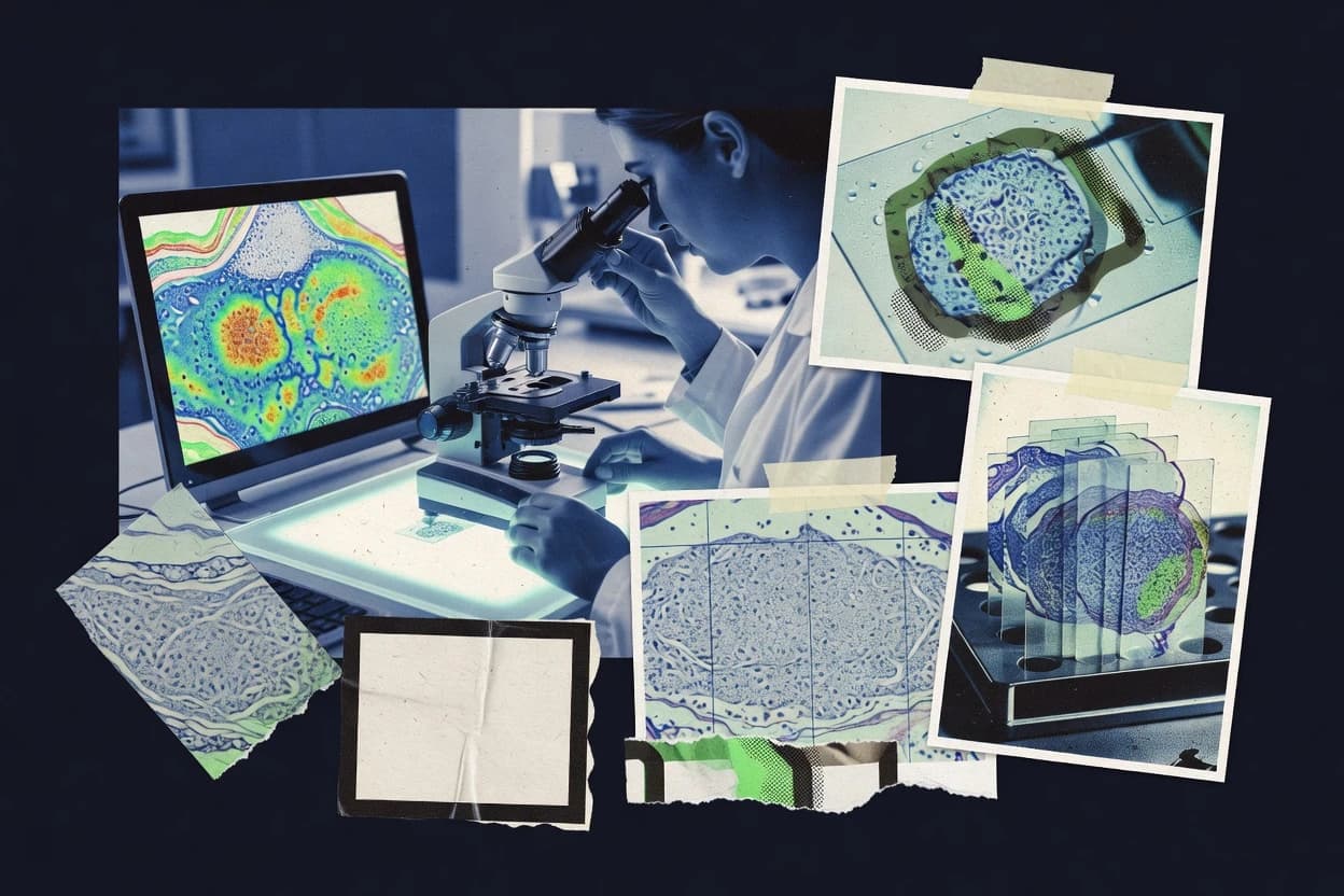
Biotechnology PharmaceuticalsTop 10 Best Histology Image Analysis Software of 2026
Top 10 Histology Image Analysis Software options ranked and compared. Review VISIOPHARM, QuPath, and CellProfiler picks for faster results.

How we ranked these tools
Core product claims cross-referenced against official documentation, changelogs, and independent technical reviews.
Analyzed video reviews and hundreds of written evaluations to capture real-world user experiences with each tool.
AI persona simulations modeled how different user types would experience each tool across common use cases and workflows.
Final rankings reviewed and approved by our editorial team with authority to override AI-generated scores based on domain expertise.
Score: Features 40% · Ease 30% · Value 30%
Gitnux may earn a commission through links on this page — this does not influence rankings. Editorial policy
Editor’s top 3 picks
Three quick recommendations before you dive into the full comparison below — each one leads on a different dimension.
VISIOPHARM
Interactive segmentation and quantification workflows for whole-slide histology images
Built for research teams quantifying biomarkers from whole-slide histology at scale.
QuPath Extensions
Editor pickCommunity plugin framework for adding segmentation and quantification tools inside QuPath
Built for teams needing specialized QuPath plugins for whole-slide histology quantification.
CellProfiler
Editor pickCellProfiler pipelines with configurable segmentation and measurement modules plus batch execution
Built for teams needing reproducible, rule-based histology quantification across batches.
Related reading
Comparison Table
This comparison table reviews histology image analysis software across core workflows such as image viewing, segmentation, quantitative measurements, and batch processing. It contrasts major tools including VISIOPHARM, QuPath Extensions, CellProfiler, Indica Labs HALO, and Aperio ImageScope to show how each option fits typical study pipelines and data formats. Readers can use the table to identify differences in analysis capabilities, automation features, and integration paths before selecting a platform.
VISIOPHARM
pathology quantificationDigital pathology image analysis platform that quantifies histology and IHC biomarkers with algorithm libraries and scoring pipelines.
Interactive segmentation and quantification workflows for whole-slide histology images
VISIOPHARM distinguishes itself with workflow-focused histology image analysis that targets quantitative tissue assessment from whole-slide images. It supports interactive and scripted analysis pipelines, including stain-aware segmentation, measurement generation, and batch processing across large slide sets. Outputs include spatial and intensity-based quantifications suitable for biomarker studies and research reporting. Extensive integration around tissue structures and marker quantification enables repeatable analysis across cohorts.
- +Whole-slide image quantification with consistent, repeatable tissue measurements
- +Stain-aware segmentation for reliable region and marker analysis
- +Batch processing for high-throughput cohorts
- +Spatial quantification that supports tissue architecture analysis
- +Workflow tooling for building and reusing analysis pipelines
- –Requires configuration effort for new stains and tissue types
- –Interactive setup can be time-consuming for large rule changes
- –Limited suitability for casual, one-off image labeling tasks
- –Custom pipelines may need technical oversight for validation
- –Complex analyses can slow down iterative parameter tuning
Best for: Research teams quantifying biomarkers from whole-slide histology at scale
More related reading
QuPath Extensions
extension ecosystemCommunity-maintained modules for QuPath that add segmentation, feature extraction, and analysis capabilities for histology workflows.
Community plugin framework for adding segmentation and quantification tools inside QuPath
QuPath Extensions adds community-built plugins to QuPath for histology and whole-slide image workflows. The extension ecosystem supports added segmentation, quantification, registration, and visualization steps beyond the base QuPath feature set. It fits labs that need specialized image analysis tasks while staying within QuPath’s tiling, ROI, and batch-processing workflow. Reproducibility improves when pipelines are driven through scripted or parameterized plugin tools.
- +Extends QuPath with targeted plugins for segmentation and quantification workflows
- +Works directly with QuPath ROIs and whole-slide tiling workflow
- +Enables specialized analysis tasks without rewriting core image handling
- +Supports reproducible batch processing through repeatable plugin parameters
- –Plugin quality varies across community contributions
- –Some extensions require manual tuning for staining and scanner variability
- –Compatibility can break after QuPath or dependency updates
- –Debugging analysis issues is harder when plugin internals are complex
Best for: Teams needing specialized QuPath plugins for whole-slide histology quantification
CellProfiler
open-source quantificationOpen-source image analysis software for high-throughput quantification of cells and features from microscopy and histology-adjacent images.
CellProfiler pipelines with configurable segmentation and measurement modules plus batch execution
CellProfiler distinguishes itself with rule-based, reproducible image analysis pipelines for histology and microscopy, built around segmentation and quantification. It supports batch processing of large image sets and exports structured measurements for downstream statistics. Core modules handle cell and nuclear segmentation, feature extraction, and intensity and morphology measurements across channels. Results integrate with workflows through overlays, CSV exports, and scripts for extending analysis logic.
- +Pipeline-based workflow for repeatable segmentation and quantification
- +Robust module library for nuclei, cells, and feature extraction
- +Batch processing supports high-throughput histology experiments
- +Exports quantitative measurements for downstream statistical analysis
- +Programmable customization through analysis scripts
- –Complex pipelines require careful parameter tuning for reliable segmentation
- –GUI-based setup can be slower than fully scripted alternatives
- –Large projects demand disciplined organization of measurements and metadata
- –Training-like accuracy improvements are limited for challenging stain variation
Best for: Teams needing reproducible, rule-based histology quantification across batches
Indica Labs HALO
quant pathologyQuantitative pathology software enables automated slide scoring, cell counting, and biomarker analysis with support for AI-assisted pipelines.
Tissue compartment and cell-level quantification on whole-slide images
HALO by Indica Labs stands out for its end-to-end histology image analysis workflow built around whole-slide processing and tissue-aware outputs. The software supports spatial tissue mapping, automated cell segmentation, and quantification across large image sets. It also enables analysis pipelines using training and rules to standardize scoring and biomarker readouts across studies. HALO’s focus on reproducible digital pathology measurements makes it a strong fit for both research studies and assay development.
- +Whole-slide analysis workflow designed for large histology image batches
- +Automated tissue compartment mapping with quantification-ready outputs
- +Cell segmentation and biomarker scoring workflows for consistent measurement
- +Pipeline-based processing reduces manual variation across analysts
- –High dataset complexity can require significant parameter tuning
- –Workflow setup takes histology-specific annotation and training effort
- –Export customization for downstream tooling can be limited by fixed formats
Best for: Teams needing automated histology quantification and reproducible scoring workflows
Aperio ImageScope
WSI viewerWhole slide image viewing and interactive pathology workflows for slide navigation, measurement, and analysis support in histology and digital pathology pipelines.
Comprehensive whole-slide visualization with annotation and quantitative measurement overlay support
Aperio ImageScope stands out with a classic whole-slide viewer and annotation workflow built for pathology use. It supports offline viewing of high-resolution slide images with fast pan, zoom, and multilayer navigation. Segmentation and analysis are enabled through application-specific algorithms delivered via companion tools and scripted workflows. Results can be stored as measurements, overlays, and reporting outputs for review and quality assurance.
- +High-resolution whole-slide viewing with responsive pan and zoom
- +Rich annotation tools for pathologist review and collaboration
- +Supports analysis overlays with quantitative measurement outputs
- +Workflow-friendly slide navigation for dense tissue regions
- +Integrates analysis results into review-ready visual layers
- –Limited evidence of fully self-contained segmentation without add-on tools
- –Automation and batch analysis depend on workflow components outside the viewer
- –Large projects can require careful system tuning for responsiveness
- –Advanced analytics need predefined algorithms and parameter setup
Best for: Pathology teams needing fast slide review with measurement overlays
HALO
WSI analysisDigital pathology software for whole slide image analysis with module-based algorithms for tissue quantification and biomarker scoring.
HALO Image Analysis software’s tissue and cell segmentation for automated scoring
HALO provides histology image analysis focused on whole-slide workflows, from tissue scanning to quantitative outputs. The software supports multi-channel analysis for common stains and enables robust region and feature detection for downstream measurements. HALO is built for repeatable pipelines with automated segmentation, scoring, and batch processing across large slide cohorts. The tool also supports integrations for managing analysis outputs and visual review of results.
- +Whole-slide analysis with automated region detection
- +Multi-channel workflows for common histology stain types
- +Batch processing for consistent scoring across large cohorts
- +Interactive visualization to validate segmentation and measurements
- +Pipeline templates support repeatable analysis across projects
- –Advanced customization often requires workflow design and expertise
- –Resource demands can be high for very large slide batches
- –Model tuning for niche markers may take iterative refinement
- –Cohort-level governance depends on external data management
- –Export formats can require additional setup for specific systems
Best for: Teams running repeatable histology scoring pipelines on large slide sets
Fiji (ImageJ Distribution)
image analysisExtensible microscopy image analysis platform with tools for segmentation, measurement, and batch processing of histology images.
Comprehensive ImageJ plugin ecosystem for segmentation, registration, and measurement in one tool
Fiji builds on ImageJ and targets image analysis workflows for microscopy, including histology staining and whole-slide style datasets. It ships with a large plugin ecosystem for segmentation, registration, and measurement, enabling end-to-end histology quantification without separate tooling. Core functionality includes interactive tools for thresholding, region-of-interest measurements, and batch processing to apply the same pipeline across many images. Fiji also supports common microscopy data formats and can be extended with additional ImageJ plugins for specialized staining and downstream analysis.
- +Plugin library covers segmentation, registration, and microscopy-specific processing tasks
- +Interactive ROI tools enable fast histology measurements from stained sections
- +Batch processing supports reproducible analysis across large image sets
- +ImageJ-based scripting and macros automate repetitive quantification steps
- –Many capabilities depend on installing and maintaining the right plugins
- –Usability can vary by plugin and may require workflow tuning
- –Performance may lag on very large whole-slide images without preprocessing
Best for: Histology groups needing plugin-driven microscopy quantification with ImageJ workflows
DeepCell
segmentationDeep learning-based segmentation toolset for cells and nuclei that supports quantitative analysis of microscopy and histology images.
Automated nucleus segmentation producing cell instance masks for per-cell quantitative measurements
DeepCell focuses on nucleus-level analysis for histology images using deep learning workflows centered on cell segmentation and object classification. The platform supports automated extraction of cell instances and morphology features from microscopy data. DeepCell also enables downstream quantification by generating per-cell outputs that can be used for biological discovery and pathology studies. Model execution is built around well-defined computer vision tasks rather than manual annotation alone.
- +Strong nucleus segmentation outputs from complex histology images
- +Provides per-cell measurements suitable for quantitative analysis pipelines
- +Deep learning workflows reduce manual annotation effort
- +Task-focused outputs support consistent morphology quantification
- –Performance can drop when staining differs from training domains
- –Less suitable for non-nuclear structures without extra modeling steps
- –Requires careful preprocessing for optimal segmentation quality
- –Limited interactive tooling for complex expert review workflows
Best for: Teams needing automated nucleus segmentation and per-cell quantification from histology images
Nikon NIS-Elements
microscopy softwareMicroscopy acquisition and image analysis software that includes measurement tools and analysis workflows for histology data.
Integrated calibration and measurement tools designed for repeatable morphometry on tissue images
Nikon NIS-Elements stands out with a microscopy-first workflow that covers acquisition, calibration, and analysis for histology and related imaging. The software provides measurement, segmentation, and morphometry tools tailored for tissue feature quantification, supported by image processing filters and batch-capable analysis. Metrology-oriented calibration tools help standardize measurements across sessions and instruments. Integrated visualization and results export support traceable quantitative outputs for tissue studies.
- +Microscopy-oriented workflow links acquisition and quantitative histology measurements
- +Calibration tools support consistent spatial measurements across datasets
- +Segmentation and morphometry features target tissue structure quantification
- –Requires strong image setup discipline for reliable segmentation results
- –Advanced workflows can feel heavy for simple stain analysis tasks
- –Integration outside Nikon microscopy ecosystems is more limited
Best for: Histology labs needing metrology-grade measurements in microscopy workflows
3DHISTECH Case Center
WSI platformWhole slide imaging and analysis workflow software that supports annotation and automated analysis tasks for histology slides.
Integrated case management linking whole-slide viewing, annotations, and analysis results
3DHISTECH Case Center stands out for its histology case-centric workflow around whole slide images and multi-level annotations. Core capabilities include digital slide viewing, annotation management, and tissue or cell analysis workflows built for pathology datasets. The tool supports collaborative review by keeping case organization, slide metadata, and analysis outputs linked. Imaging analysis tasks can be exported as measurable results tied to the original slide context for downstream reporting.
- +Case-first organization keeps annotations and results tied to the same slide context
- +Whole slide visualization supports detailed navigation for morphology review
- +Analysis outputs remain connected to case and metadata for traceable documentation
- –Workflow is oriented to case management rather than lightweight batch scripting
- –Advanced analysis setup can require careful parameter tuning per stain and morphology
- –Export and integration paths can be limiting for highly customized pipelines
Best for: Pathology teams organizing annotated whole-slide studies with traceable analysis outputs
How to Choose the Right Histology Image Analysis Software
This buyer's guide explains how to select histology image analysis software for biomarker quantification, cell or nucleus segmentation, and whole-slide scoring workflows. It covers VISIOPHARM, QuPath Extensions, CellProfiler, Indica Labs HALO, Aperio ImageScope, HALO by akoya, Fiji (ImageJ Distribution), DeepCell, Nikon NIS-Elements, and 3DHISTECH Case Center. Each section ties selection criteria to concrete capabilities such as stain-aware segmentation in VISIOPHARM and nucleus instance masks in DeepCell.
What Is Histology Image Analysis Software?
Histology image analysis software turns stained tissue images into quantified measurements such as tissue compartment fractions, marker intensity, nuclei or cells counts, and morphometry features. The software targets common pain points like repeatability across cohorts, batch processing of large slide sets, and consistent segmentation from whole-slide images. Some tools are built for interactive pathology review with annotation and measurement overlays like Aperio ImageScope. Other tools focus on automated, pipeline-driven quantification like CellProfiler and VISIOPHARM.
Key Features to Look For
These features determine whether a tool can deliver repeatable quantitative outputs from your specific histology stains, image sizes, and workflow constraints.
Whole-slide quantification with spatial and intensity outputs
VISIOPHARM supports whole-slide image quantification with spatial measurements and intensity-based outputs. Indica Labs HALO provides tissue compartment mapping with quantification-ready results for biomarker scoring workflows.
Stain-aware segmentation and tissue-aware region detection
VISIOPHARM uses stain-aware segmentation to generate consistent region and marker measurements across whole-slide images. HALO by akoya also supports tissue and cell segmentation designed for automated scoring in repeatable pipelines.
Batch processing built for large cohorts
CellProfiler supports batch execution of reproducible segmentation and feature extraction across large image sets and exports structured measurements. VISIOPHARM and Indica Labs HALO both provide batch workflows across large slide cohorts designed to reduce manual variation.
Configurable pipeline frameworks for reproducible measurement logic
CellProfiler centers on rule-based pipelines built from configurable segmentation and quantification modules. VISIOPHARM offers interactive and scripted analysis pipelines that can be reused for consistent measurement generation.
Deep learning instance segmentation for per-cell or per-nucleus quantification
DeepCell delivers automated nucleus segmentation that outputs cell instance masks for per-cell quantitative measurements. This is the most direct fit when the analysis output must be object-level instance masks rather than only region-level scoring.
Case management and traceable outputs linked to slide context
3DHISTECH Case Center ties whole-slide viewing, annotations, and analysis outputs to case organization and slide metadata for traceable documentation. Aperio ImageScope supports review-ready visual layers by storing measurements and overlays tied to pathology workflows.
How to Choose the Right Histology Image Analysis Software
Selection should follow the target output type, the required workflow automation level, and the needed integration of viewing and measurement logic.
Match the tool to the required output unit: slide-level, tissue-compartment, or per-cell instances
For slide-level biomarker quantification with spatial architecture outputs, VISIOPHARM provides whole-slide measurements that combine intensity and spatial quantification. For tissue compartment scores and cell-level quantification across cohorts, Indica Labs HALO and HALO by akoya both focus on tissue-aware mapping plus cell segmentation for scoring.
Choose the segmentation approach based on stain variability and structure complexity
When segmentation must adapt to stains to support reliable region and marker analysis, VISIOPHARM’s stain-aware segmentation is designed for consistent measurement generation. For nucleus-focused workflows where per-cell outputs require instance masks, DeepCell provides automated nucleus segmentation built around deep learning and per-cell morphology extraction.
Decide how automation should be built and governed across analysts
For rule-based, repeatable measurement pipelines that can be parameterized for consistent batch runs, CellProfiler offers configurable modules for nuclei, cells, intensity, and morphology measurements plus CSV exports. For teams that need to stay in a QuPath environment while adding specialized segmentation and quantification tasks, QuPath Extensions extends QuPath’s tiling, ROI, and batch workflow through community plugins.
Plan for setup and maintenance workload before committing to complex pipelines
VISIOPHARM can require configuration effort when introducing new stains and tissue types and complex analyses can slow iterative parameter tuning. Indica Labs HALO can require significant parameter tuning for high dataset complexity and workflow setup can take histology-specific annotation and training effort.
If pathology review, annotation, and traceability are central, prioritize integrated case and visualization tools
When the workflow must center on whole-slide review with annotation and measurement overlays, Aperio ImageScope provides responsive pan and zoom plus rich annotation and overlay outputs. When case organization and traceability between annotations, metadata, and results are required, 3DHISTECH Case Center keeps slide context tightly linked to measurable outputs.
Who Needs Histology Image Analysis Software?
Different teams need different output formats and workflow controls, from whole-slide biomarker quantification to per-cell nucleus instance masks and case-linked traceable outputs.
Research teams quantifying biomarkers from whole-slide histology at scale
VISIOPHARM is the best fit for whole-slide image quantification with stain-aware segmentation, spatial quantification, batch processing, and reusable workflow tooling. Indica Labs HALO also fits teams that need automated tissue compartment mapping and reproducible cell segmentation for scoring across large slide sets.
Teams that rely on QuPath for their pathology image workflow but need added segmentation and quantification capabilities
QuPath Extensions is designed to add community-built plugins for segmentation, quantification, registration, and visualization inside QuPath. This choice supports reproducible batch processing through repeatable plugin parameters tied to QuPath’s ROI and whole-slide tiling workflow.
Teams that need rule-based, reproducible segmentation and measurement pipelines across many experiments
CellProfiler excels for pipeline-based segmentation and quantification of cells and features with batch processing and structured measurement exports. Fiji (ImageJ Distribution) fits teams that want plugin-driven microscopy quantification with ImageJ scripting and batch processing when the team can manage plugin installation and workflow tuning.
Teams focused on nuclei and per-cell morphometry outputs rather than only region scoring
DeepCell is built for nucleus-level analysis and produces cell instance masks and per-cell measurements for quantitative pipelines. This is the strongest match when downstream analytics require object-level instance segmentation outputs for each nucleus.
Common Mistakes to Avoid
Several recurrent pitfalls show up across these tools, and avoiding them prevents wasted implementation cycles and inconsistent quantitative outputs.
Choosing a viewer-first workflow when automated quantification pipelines are required
Aperio ImageScope is optimized for whole-slide viewing, annotation, and measurement overlays, while automation depends on application-specific algorithms delivered via companion tools and scripted workflows. Teams needing automated scoring and batch quantification should prioritize VISIOPHARM, Indica Labs HALO, or HALO by akoya.
Underestimating stain and tissue model setup work for complex pipelines
VISIOPHARM can require configuration effort for new stains and tissue types, and complex analyses can slow iterative parameter tuning. Indica Labs HALO can require significant parameter tuning and histology-specific annotation and training effort when dataset complexity is high.
Relying on community plugins without a governance plan for validation and maintenance
QuPath Extensions can break compatibility after QuPath or dependency updates and plugin quality can vary across community contributions. Teams that select QuPath Extensions should allocate time for validation because debugging plugin internals can be harder.
Trying to segment non-nuclear structures with a nucleus-first tool without extra modeling steps
DeepCell is focused on nucleus-level analysis and is less suitable for non-nuclear structures without additional modeling steps. For mixed tissue structures and broader biomarker region analysis, VISIOPHARM, Indica Labs HALO, or HALO by akoya are built for tissue and compartment quantification.
How We Selected and Ranked These Tools
we evaluated VISIOPHARM, QuPath Extensions, CellProfiler, Indica Labs HALO, Aperio ImageScope, HALO by akoya, Fiji (ImageJ Distribution), DeepCell, Nikon NIS-Elements, and 3DHISTECH Case Center on three sub-dimensions. Features received weight 0.4, ease of use received weight 0.3, and value received weight 0.3. Overall equals 0.40 × features plus 0.30 × ease of use plus 0.30 × value. VISIOPHARM separated itself by combining stain-aware whole-slide segmentation with workflow tooling for interactive and scripted quantification pipelines, which scored strongly in features and also supported repeatable batch measurement workflows.
Frequently Asked Questions About Histology Image Analysis Software
Which tools are best for quantitative biomarker measurement from whole-slide histology images?
What options exist for standardized, reproducible histology analysis across large slide cohorts?
How do plugin-based workflows compare with end-to-end histology analysis platforms?
Which software is most suitable for tissue compartment analysis and spatial mapping?
Which tools focus on nucleus or cell-level segmentation using deep learning?
What is the best fit for fast whole-slide review and measurement overlay workflows?
How do imaging metrology and calibration capabilities affect histology measurements?
Which platforms support scripted or pipeline-driven execution rather than fully manual annotation?
What are common technical bottlenecks when analyzing whole-slide images, and how do tools address them?
How should teams decide between case management and analysis-first tools?
Conclusion
After evaluating 10 biotechnology pharmaceuticals, VISIOPHARM stands out as our overall top pick — it scored highest across our combined criteria of features, ease of use, and value, which is why it sits at #1 in the rankings above.
Use the comparison table and detailed reviews above to validate the fit against your own requirements before committing to a tool.
Tools reviewed
Primary sources checked during evaluation.
Referenced in the comparison table and product reviews above.
Keep exploring
Comparing two specific tools?
Software Alternatives
See head-to-head software comparisons with feature breakdowns, pricing, and our recommendation for each use case.
Explore software alternatives→In this category
Biotechnology Pharmaceuticals alternatives
See side-by-side comparisons of biotechnology pharmaceuticals tools and pick the right one for your stack.
Compare biotechnology pharmaceuticals tools→FOR SOFTWARE VENDORS
Not on this list? Let’s fix that.
Our best-of pages are how many teams discover and compare tools in this space. If you think your product belongs in this lineup, we’d like to hear from you—we’ll walk you through fit and what an editorial entry looks like.
Apply for a ListingWHAT THIS INCLUDES
Where buyers compare
Readers come to these pages to shortlist software—your product shows up in that moment, not in a random sidebar.
Editorial write-up
We describe your product in our own words and check the facts before anything goes live.
On-page brand presence
You appear in the roundup the same way as other tools we cover: name, positioning, and a clear next step for readers who want to learn more.
Kept up to date
We refresh lists on a regular rhythm so the category page stays useful as products and pricing change.