GITNUXSOFTWARE ADVICE
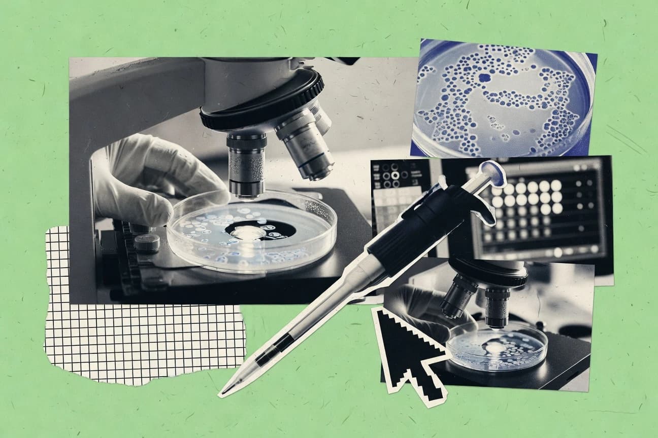
Biotechnology PharmaceuticalsTop 10 Best Cell Counting Software of 2026
Compare the top Cell Counting Software options in a ranking of best tools, including ImageJ, Fiji, and CellProfiler. Explore picks.

How we ranked these tools
Core product claims cross-referenced against official documentation, changelogs, and independent technical reviews.
Analyzed video reviews and hundreds of written evaluations to capture real-world user experiences with each tool.
AI persona simulations modeled how different user types would experience each tool across common use cases and workflows.
Final rankings reviewed and approved by our editorial team with authority to override AI-generated scores based on domain expertise.
Score: Features 40% · Ease 30% · Value 30%
Gitnux may earn a commission through links on this page — this does not influence rankings. Editorial policy
Editor’s top 3 picks
Three quick recommendations before you dive into the full comparison below — each one leads on a different dimension.
ImageJ
Watershed-based particle separation with thresholding and size filtering
Built for researchers needing customizable, reproducible microscopy cell counting workflows.
Fiji
Editor pickWatershed-based instance segmentation for separating touching cells
Built for labs needing customizable microscopy cell counting with automation.
CellProfiler
Editor pickPipeline-based object segmentation and measurement across batches using CellProfiler modules
Built for research teams needing configurable microscopy cell counting with reproducible pipelines.
Related reading
Comparison Table
This comparison table evaluates cell counting software used for image-based segmentation, nucleus detection, and quantitative output across microscopy workflows. It contrasts ImageJ and Fiji with automation-focused tools like CellProfiler and QuPath, plus instrument-driven options such as IN Cell Analyzer, highlighting differences in setup, analysis pipeline control, and export of cell metrics. Readers can use the table to match tool capabilities to dataset types, imaging formats, and desired outputs like counts per region, size distributions, and marker-specific labeling.
ImageJ
open-sourceOpen-source image analysis software that supports cell counting workflows through built-in tools and plug-ins such as Simple Neurite Tracer, Cell Counter, and Trainable Weka Segmentation.
Watershed-based particle separation with thresholding and size filtering
ImageJ stands out with its open-source image analysis engine and long-standing community of plugins for microscopy and biological workflows. For cell counting, it supports thresholding, watershed-based separation of touching objects, and measurements tied to calibrated image scales. Its extensibility enables custom segmentation and counting pipelines through macros, scripts, and plugin modules, including batch processing over folders of images.
- +Watershed segmentation separates touching cells for counting workflows.
- +Calibrated measurements connect counts to area, size, and spatial scale.
- +Macro and plugin ecosystem enables reusable pipelines across experiments.
- +Batch processing supports consistent counting on large image sets.
- –Setup and parameter tuning require image-specific trial and error.
- –User interface can feel technical for fully guided cell counting.
- –Advanced automation often needs scripting or third-party plugins.
Best for: Researchers needing customizable, reproducible microscopy cell counting workflows
More related reading
Fiji
scientific imagingDistribution of ImageJ curated for scientific imaging that includes cell-counting and segmentation workflows like watershed-based counting and machine-learning segmentation via Trainable Weka.
Watershed-based instance segmentation for separating touching cells
Fiji stands out as an extensible image analysis platform built on ImageJ with strong community-driven plugins for microscopy tasks. It delivers cell counting workflows through thresholding, watershed segmentation, and batch processing that can be automated with recorded macros.
High accuracy is achievable for well-formed microscopy images, especially when custom segmentation steps are needed. It also supports reproducible analysis via scriptable pipelines and consistent measurement outputs.
- +Large plugin ecosystem for segmentation and specialized cell counting
- +Scriptable macros and batch processing support repeatable experiments
- +Powerful segmentation tools like watershed and customizable thresholds
- +Rich measurement outputs export cleanly to downstream analysis
- +Active community support and documented workflows
- –Segmentation quality depends heavily on image preprocessing choices
- –Complex workflows require scripting or tuning beyond basic clicks
- –No single unified counting wizard for all microscopy styles
- –Large datasets can slow down without careful optimization
Best for: Labs needing customizable microscopy cell counting with automation
CellProfiler
high-content analysisOpen-source platform for high-content image analysis that performs segmentation and quantitative cell counting using pipelines for microscopy datasets.
Pipeline-based object segmentation and measurement across batches using CellProfiler modules
CellProfiler stands out for turning microscopy image analysis into reproducible, scriptable workflows using a point-and-click interface. It supports segmentation of cells, nuclei, and other structures and then measures features for each detected object.
Outputs can drive quantitative cell counting, morphology metrics, and downstream statistics across large image batches. Strong batch pipelines and modular modules make it suitable for complex assays, including multi-channel experiments.
- +Modular image analysis pipelines enable end-to-end cell counting workflows
- +Robust segmentation supports nuclei, cells, and region-based measurements
- +Batch processing handles large datasets with consistent measurement outputs
- +Outputs integrate with downstream tools via exported measurements tables
- +Reproducibility improves through saved pipelines and configurable parameters
- –Segmentation tuning often requires iterative parameter adjustments for new samples
- –Scaling to very large cohorts can demand careful workflow and compute planning
- –Advanced custom logic requires pipeline edits beyond simple point-and-click usage
- –User setup complexity can slow initial adoption versus simpler counters
Best for: Research teams needing configurable microscopy cell counting with reproducible pipelines
More related reading
QuPath
digital pathologyOpen-source digital pathology software that counts cells and analyzes biomarker-stained tissue using detection, segmentation, and scripting workflows.
QP scripting and machine-learning-assisted cell detection workflows
QuPath stands out for using a Java-based, scriptable image analysis workflow for whole-slide and multiplexed tissue images. Core cell counting capabilities include segmentation, detection, and class-based counting with measurement export for downstream analysis. It also supports batch processing through scripting to standardize counting across large slide sets.
- +Scriptable workflow enables reproducible segmentation and counting across many slides
- +Flexible detection and segmentation supports diverse stain and morphology workflows
- +Measurement and annotation exports support downstream statistics and QC
- +Batch processing lets large cohorts be analyzed with consistent parameters
- –Setup and tuning for accurate segmentation often requires expertise
- –Workflow scaling depends on hardware and memory for large whole-slide images
- –Graphical configuration can be slower than pure code pipelines for specialists
Best for: Labs needing reproducible whole-slide cell counting with programmable workflows
IN Cell Analyzer
instrument-integratedInstrument-integrated imaging and analysis software from Cytiva that supports cell counting and quantitative phenotyping for automated microscopy.
Built-in high-content image analysis for automated cell counting and quantification
IN Cell Analyzer is a Cytiva imaging-and-analysis solution designed for automated cell counting and related cellular measurements from microscopy outputs. It supports high-content workflows with plate-compatible acquisition, image-based segmentation, and quantified outputs suitable for screening and assay development.
The product also emphasizes traceable analysis steps through data management for experiments that produce large image sets. It is best suited to teams standardizing visual assays where repeatable counts across conditions matter more than ad hoc customization.
- +Automates image segmentation and cell counting across large plate experiments
- +High-content outputs support downstream metrics beyond simple counts
- +Workflow traceability helps keep analysis consistent across runs
- –Best results depend on careful image and segmentation setup
- –Deep tuning and advanced customization require specialist configuration
Best for: Biology teams running high-content microscopy cell counting at scale
MetaMorph
microscopy analysisMicroscopy acquisition and analysis platform from Molecular Devices that supports image processing steps used for cell counting pipelines.
Measurement-based object identification for counting cells after customizable image preprocessing
MetaMorph stands out for coupling cell imaging workflows with analysis tools in a single desktop environment from Molecular Devices. It supports quantitative cell counting by combining image acquisition, preprocessing, and measurement-driven object identification.
The software is designed for microscopy-centric labs that need reproducible counts across experiments and plates. It also supports scripting and method customization for specialized segmentation and counting rules.
- +Integrated microscopy workflow links acquisition and quantification steps
- +Configurable segmentation enables consistent cell counting across varied images
- +Batch processing supports throughput for multiwell plates and experiments
- –Segmentation tuning can require technical familiarity with image processing
- –Graphical setup can feel complex for simple one-off counting tasks
- –Scaling bespoke counting logic often benefits from scripting expertise
Best for: Microscopy teams needing methodized, reproducible cell counting with custom segmentation
More related reading
Visiopharm
enterprise pathologyCommercial image analysis software suite that counts cells in histology and tissue images using trained segmentation and quantification modules.
Visiopharm image analysis pipelines with reviewable overlays for validation of counted cells
Visiopharm stands out by combining microscopy-focused cell counting with configurable analysis workflows built for research and imaging labs. The platform supports automated object detection, segmentation, and measurement on microscopic images to reduce manual counting variance.
It also emphasizes repeatable analysis through template-based pipelines and reviewable outputs that can be audited against raw images. The core value is dependable quantification for heterogeneous samples where cell boundaries and image quality vary.
- +Flexible segmentation and cell detection workflows for complex microscopy images
- +Configurable analysis pipelines support repeatable counting across experiments
- +Overlay outputs make it easier to validate counts against original imagery
- –Setup and tuning require image-analytics expertise for reliable segmentation
- –Workflow configuration can feel heavy for simple batch counting tasks
- –Integration paths and deployment specifics can add friction in mixed lab environments
Best for: Labs needing configurable microscopy cell counting with auditable image overlays
Definiens Developer XD
enterprise informaticsEnterprise image informatics software that enables cell and tissue quantification from whole-slide and microscopy images with rule- and AI-based analysis.
Definiens Developer XD multi-step rule-based and machine learning image classification for cell phenotyping
Definiens Developer XD stands out for turning image analysis design into a programmable workflow for microscopy cell counting and phenotype measurement. The platform supports rule-based and machine learning classification pipelines that operate on single-cell and multi-cell image datasets. It also includes tools for annotating training data, validating segmentation and counting results, and exporting measurements for downstream analysis.
- +Configurable image analysis pipelines for robust cell segmentation and counting
- +Integrated training and classification tools for cell phenotype measurement
- +Validation-focused workflow to check segmentation and counting accuracy
- +Export-ready measurements for downstream statistics and reporting
- –Workflow authoring can feel engineering-heavy for non-developers
- –Result quality depends strongly on image acquisition consistency
- –Large project setup and tuning can slow first deployments
Best for: Research groups needing configurable, classification-driven cell counting workflows
More related reading
Imaris
3D microscopy3D microscopy visualization and analysis software that detects and counts cells and nuclei in volumetric datasets using segmentation and tracking features.
Surface and spot-based segmentation for automated 3D cell and nucleus counting
Imaris stands out for counting cells inside 3D and time-series microscopy data using interactive visualization plus analysis. It provides automated segmentation and object counting with configurable parameters for nuclei, cells, and other structures. Researchers can generate measurements, track populations over time, and export results for downstream statistics and reporting.
- +3D and time-lapse cell counting with segmentation tuned to imaging noise
- +Automated object detection produces counts, volumes, and intensity metrics
- +Object tracking supports studying population changes across timepoints
- +Interactive refinement makes it practical to correct segmentation errors
- +Exports object tables for direct downstream analysis workflows
- –Parameter tuning can be time-consuming for unfamiliar microscope settings
- –Performance depends heavily on dataset size and hardware capacity
- –Workflow setup requires more training than 2D-only counting tools
Best for: Teams quantifying 3D and time-lapse cell populations with segmentation-heavy workflows
CellProfiler Analyst
data reviewOptional analysis and visualization layer for CellProfiler outputs that supports counting-related quality control and review of quantitative results.
Population gating and filtering over extracted CellProfiler measurements
CellProfiler Analyst stands out by coupling CellProfiler pipeline outputs with interactive statistical exploration for imaging experiments. It supports plate and experiment level visualization, including scatter plots, histograms, box plots, and heatmaps.
The tool enables filtering and gating across images using computed features to support cell counting workflows. Data can be exported for downstream statistical analysis and reporting.
- +Interactive plots support rapid QC of cell measurements across images
- +Filtering and gating make it easier to refine cell populations
- +Exports processed measurements for downstream statistics and reporting
- –Relies on CellProfiler preprocessing, so setup is multi-step
- –Feature selection and gating workflows require familiarization
- –Large datasets can feel slower during interactive exploration
Best for: Teams analyzing CellProfiler outputs with interactive population QC and cell counts
How to Choose the Right Cell Counting Software
This buyer's guide helps teams choose cell counting software by mapping workflow needs to concrete tool capabilities in ImageJ, Fiji, CellProfiler, QuPath, IN Cell Analyzer, MetaMorph, Visiopharm, Definiens Developer XD, Imaris, and CellProfiler Analyst. The guide covers segmentation and counting features, automation and batch handling, measurement and export outputs, and QC workflows that keep counts reliable across large microscopy datasets. Common implementation pitfalls are also translated into tool-specific avoidance tactics.
What Is Cell Counting Software?
Cell counting software segments cells or nuclei in microscopy images and then converts detected objects into measurable counts and per-object feature tables. It solves variability from manual counting by applying thresholding, watershed separation, detection logic, and configurable pipelines that produce consistent outputs across batches. Teams use these tools for whole-slide tissue quantification in QuPath, automated high-content plate workflows in IN Cell Analyzer, and 3D time-series counting in Imaris. Practical systems like CellProfiler and Fiji also support measurement exports that feed downstream statistics and reporting workflows.
Key Features to Look For
These capabilities drive count accuracy, reproducibility, and throughput for the specific microscopy styles represented by ImageJ, Fiji, CellProfiler, QuPath, IN Cell Analyzer, MetaMorph, Visiopharm, Definiens Developer XD, Imaris, and CellProfiler Analyst.
Watershed separation for touching cells
Watershed-based instance separation prevents over-merging when cells touch closely. ImageJ delivers watershed-based particle separation with thresholding and size filtering, and Fiji provides watershed-based instance segmentation tuned for separating touching cells in microscopy images.
Batch processing for large image sets and plate experiments
Batch processing keeps the same segmentation and counting logic consistent across many fields, wells, or slides. CellProfiler supports batch processing through modular pipelines, and ImageJ and Fiji support batch processing via macros and repeatable analysis steps.
Pipeline-based automation that preserves reproducibility
Automation through saved pipelines reduces manual parameter drift across experiments. CellProfiler runs segmentation and measurement as saved pipelines using configurable modules, while QuPath uses scriptable workflows to standardize detection and counting across many slides.
Rule-based and machine learning classification for phenotype-aware counting
Classification-driven counting supports cell phenotyping rather than counting one object class only. QuPath combines scripting with machine-learning-assisted cell detection, and Definiens Developer XD adds multi-step rule-based and machine learning classification with integrated training and validation tools.
Measurement export for downstream statistics and QC
Export-ready outputs make counts actionable for analysis, gating, and reporting. Fiji produces measurement outputs that export cleanly to downstream analysis, and CellProfiler exports measurement tables that integrate with downstream statistics workflows.
Audit and QC views tied to counting outputs
QC tooling helps validate segmentation accuracy and count logic against raw imagery and computed features. Visiopharm provides overlay outputs that support validation of counted cells against original imagery, and CellProfiler Analyst enables population gating and filtering over extracted CellProfiler measurements.
How to Choose the Right Cell Counting Software
The right choice matches the imaging modality and workflow style to the tool’s segmentation, automation, measurement, and QC capabilities.
Match the modality and image type to segmentation capabilities
For 2D microscopy where touching cells must be separated, ImageJ and Fiji fit well because both support thresholding plus watershed-based instance separation with size filtering. For whole-slide tissue with class-based counting needs, QuPath supports detection, segmentation, and class-based counting in scriptable workflows. For 3D and time-lapse datasets, Imaris is built for automated object counting with segmentation tuned to imaging noise and object tracking across timepoints.
Decide how much automation and reproducibility must be baked into the workflow
For end-to-end reproducible pipelines across large microscopy batches, CellProfiler excels with modular modules that run segmentation and measurements consistently. For scripted and programmable slide standardization, QuPath uses scripting workflows and supports batch processing through standardized parameters. For controlled high-content screening across plate experiments, IN Cell Analyzer provides built-in high-content image analysis that automates segmentation and cell counting at scale.
Choose how segmentation and object detection logic will be authored
If customizable and reusable analysis logic is required, ImageJ and Fiji support macros, scripts, and a plugin ecosystem that enables custom segmentation pipelines. If methodized lab workflows must link acquisition to measurement, MetaMorph provides integrated microscopy workflow steps that combine acquisition, preprocessing, and measurement-driven object identification. If phenotype measurement depends on training data and classification, Definiens Developer XD supports rule-based and machine learning classification with training, validation, and export-ready measurements.
Plan for validation, auditability, and interactive QC
If validated overlays are needed to audit segmentation against raw imagery, Visiopharm supplies reviewable outputs with overlay views that help confirm counted cells. If population-level QC is needed after quantification, CellProfiler Analyst adds interactive statistical exploration, filtering, and gating over extracted CellProfiler measurements. If workflow traceability across runs matters for high-content studies, IN Cell Analyzer emphasizes traceable analysis steps tied to data management for large image sets.
Confirm output requirements for downstream analysis and reporting
If per-object feature tables must feed downstream analysis, Fiji and CellProfiler both generate rich measurement outputs that export into analysis-ready formats. If whole-slide annotations and exports are required for downstream statistics and QC, QuPath supports measurement and annotation exports. If exported object tables are needed for reporting and tracking population changes, Imaris exports object tables with counts plus volumes and intensity metrics.
Who Needs Cell Counting Software?
Cell counting software benefits teams that must turn microscopy images into consistent, batch-ready quantitative counts and per-object measurements.
Researchers needing customizable, reproducible 2D microscopy cell counting workflows
ImageJ and Fiji fit this use case because both support watershed-based separation with thresholding and size filtering and both can be automated through macros, plugins, and batch processing. These tools are also suitable when segmentation needs tuning for different sample preparations without losing repeatability across runs.
Research teams building configurable, reproducible pipelines for large batch microscopy assays
CellProfiler is designed for modular pipeline-based segmentation and measurement across batches with configurable modules that output measurement tables for downstream statistics. CellProfiler Analyst is the right companion when interactive population QC and gating must be performed on extracted CellProfiler features.
Labs doing whole-slide and multiplex tissue quantification with standardized slide pipelines
QuPath supports detection, segmentation, and class-based counting with measurement export for downstream statistics and QC using scriptable workflows. Definiens Developer XD fits when cell phenotyping requires multi-step rule-based and machine learning classification with training data annotation and validation.
Teams quantifying 3D and time-lapse cell populations where tracking matters
Imaris fits because it provides automated segmentation and object counting for volumetric data, plus object tracking to study population changes across timepoints. Image quality variation is handled through segmentation parameters tuned to imaging noise, with interactive refinement to correct segmentation errors.
Common Mistakes to Avoid
Several recurring pitfalls show up across tools, especially when segmentation assumptions do not match the microscope images or when QC and validation steps are skipped.
Choosing a counting workflow that cannot separate touching cells
Watershed separation is a core requirement for dense samples with touching cells, so tools without watershed instance separation increase merge errors. ImageJ and Fiji both use thresholding with watershed-based instance separation, which directly targets touching-cell over-merging.
Treating parameters as one-size-fits-all across different sample prep
Segmentation quality depends heavily on preprocessing choices and parameter tuning, which can break across new samples. Fiji and CellProfiler require iterative parameter adjustments when image preprocessing changes, so saved pipelines and consistent preprocessing steps are necessary to keep counts stable.
Skipping audit overlays or QC views for segmentation accuracy
Counting errors often come from incorrect boundaries and missed objects, which cannot be fixed by counts alone. Visiopharm provides reviewable overlay outputs for validation against raw imagery, and CellProfiler Analyst provides gating and filtering over extracted CellProfiler measurements for population-level QC.
Over-customizing without a plan for reproducibility at batch scale
Advanced automation that relies on scripting or deep customization can slow adoption if reproducibility is not planned upfront. ImageJ, Fiji, and QuPath support automation through macros, scripts, and batch processing, while CellProfiler delivers reproducible end-to-end pipelines through saved modules.
How We Selected and Ranked These Tools
we evaluated every tool on three sub-dimensions, features with weight 0.4, ease of use with weight 0.3, and value with weight 0.3. the overall rating is the weighted average of those three sub-dimensions using overall = 0.40 × features + 0.30 × ease of use + 0.30 × value. ImageJ separated itself from lower-ranked tools through concrete watershed-based particle separation combined with thresholding and size filtering, which directly strengthens segmentation quality for touching objects and improves count reliability in feature scoring. That combination of segmentation capability plus reusable macro and plugin pipelines pushed ImageJ to the top position with an overall rating that reflects its strengths across features, ease of use, and value.
Frequently Asked Questions About Cell Counting Software
Which cell counting tools handle touching cells better during segmentation?
What software is best for fully reproducible, scriptable microscopy cell counting pipelines?
Which options are suited for whole-slide and multiplexed tissue cell counting rather than single fields of view?
Which tools support high-throughput screening workflows tied to plate-based experiments?
How do researchers review and audit counted cells against the raw image data?
Which tool is strongest for 3D cell counting and time-lapse population tracking?
Which solutions fit multi-step phenotype classification rather than only counting cells?
What are common segmentation and counting failure modes across these tools and how do they get addressed?
Which approach is best for custom segmentation rules that require building a counting pipeline from components?
Conclusion
After evaluating 10 biotechnology pharmaceuticals, ImageJ stands out as our overall top pick — it scored highest across our combined criteria of features, ease of use, and value, which is why it sits at #1 in the rankings above.
Use the comparison table and detailed reviews above to validate the fit against your own requirements before committing to a tool.
Tools reviewed
Primary sources checked during evaluation.
Referenced in the comparison table and product reviews above.
Keep exploring
Comparing two specific tools?
Software Alternatives
See head-to-head software comparisons with feature breakdowns, pricing, and our recommendation for each use case.
Explore software alternatives→In this category
Biotechnology Pharmaceuticals alternatives
See side-by-side comparisons of biotechnology pharmaceuticals tools and pick the right one for your stack.
Compare biotechnology pharmaceuticals tools→FOR SOFTWARE VENDORS
Not on this list? Let’s fix that.
Our best-of pages are how many teams discover and compare tools in this space. If you think your product belongs in this lineup, we’d like to hear from you—we’ll walk you through fit and what an editorial entry looks like.
Apply for a ListingWHAT THIS INCLUDES
Where buyers compare
Readers come to these pages to shortlist software—your product shows up in that moment, not in a random sidebar.
Editorial write-up
We describe your product in our own words and check the facts before anything goes live.
On-page brand presence
You appear in the roundup the same way as other tools we cover: name, positioning, and a clear next step for readers who want to learn more.
Kept up to date
We refresh lists on a regular rhythm so the category page stays useful as products and pricing change.