GITNUXSOFTWARE ADVICE
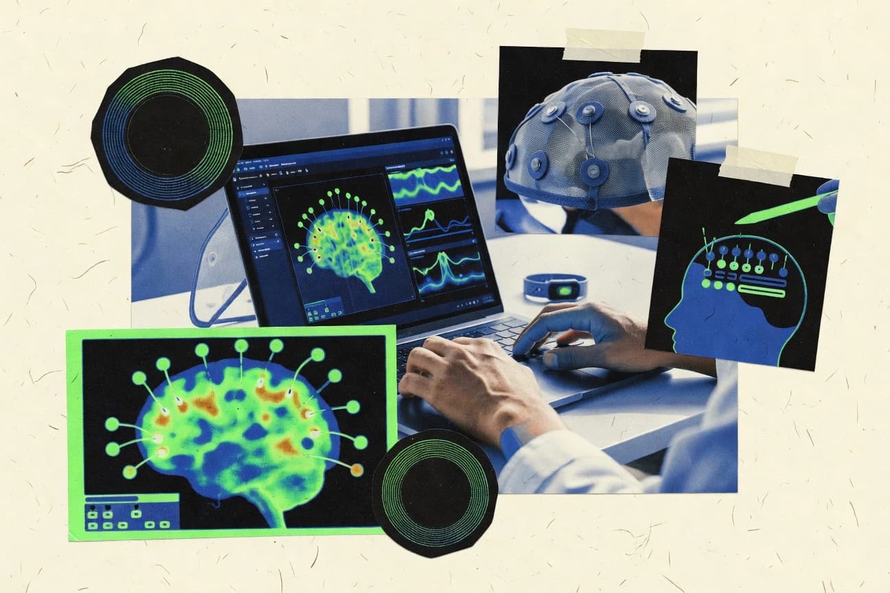
Science ResearchTop 9 Best Brainmapping Software of 2026
Top 10 Brainmapping Software picks ranked and compared for MRI workflows. Explore best tools like BrainNet Viewer, MRtrix3, and FSL.

How we ranked these tools
Core product claims cross-referenced against official documentation, changelogs, and independent technical reviews.
Analyzed video reviews and hundreds of written evaluations to capture real-world user experiences with each tool.
AI persona simulations modeled how different user types would experience each tool across common use cases and workflows.
Final rankings reviewed and approved by our editorial team with authority to override AI-generated scores based on domain expertise.
Score: Features 40% · Ease 30% · Value 30%
Gitnux may earn a commission through links on this page — this does not influence rankings. Editorial policy
Editor’s top 3 picks
Three quick recommendations before you dive into the full comparison below — each one leads on a different dimension.
BrainNet Viewer
High-control surface-based network visualization with customizable node and edge rendering
Built for researchers visualizing brain graphs with high-quality 3D figures and quick iteration.
MRtrix3
Editor pickSpherical deconvolution with multi-tissue response functions and constrained tractography
Built for research teams running diffusion MRI tractography pipelines with custom analysis control.
FSL
Editor pickNIfTI-compatible FEAT GLM pipeline for fMRI analysis with common preprocessing steps
Built for research labs needing reproducible MRI, fMRI, and diffusion processing pipelines.
Related reading
Comparison Table
This comparison table reviews widely used brain mapping and neuroimaging toolkits, including BrainNet Viewer, MRtrix3, FSL, FreeSurfer, and ANTs. It summarizes what each package can do for data preprocessing, registration and segmentation, diffusion and tractography, and volumetric or surface-based analysis. Readers can use the table to match tool capabilities to common workflows in neuroimaging research and clinical imaging pipelines.
BrainNet Viewer
connectome visualizationVisualizes brain networks and neuroimaging-derived connectivity data on 3D brain models for research figures and exploratory analysis.
High-control surface-based network visualization with customizable node and edge rendering
BrainNet Viewer stands out for fast, interactive 3D visualization of brain networks on common neuroimaging surfaces and volumes. The core workflow supports importing node and edge data, rendering networks, customizing visual styles, and exporting publication-ready figures. It also includes utilities for managing coordinate systems and for linking multiple brain views for exploratory analysis.
- +Interactive 3D network rendering on brain surfaces and volumes
- +Strong control over node and edge styling for publication figures
- +Supports common network plotting workflows without heavy infrastructure
- –Configuration often depends on correct coordinate and file formatting
- –Limited built-in analytics beyond visualization and figure generation
- –Windows-only usage patterns and setup friction for some environments
Best for: Researchers visualizing brain graphs with high-quality 3D figures and quick iteration
More related reading
MRtrix3
diffusion MRI processingPerforms diffusion MRI processing including tractography and connectome generation with command-line pipelines used in brain mapping workflows.
Spherical deconvolution with multi-tissue response functions and constrained tractography
MRtrix3 stands out for its command-line driven diffusion and structural MRI workflow, with tight integration across preprocessing, modeling, and tractography. It supports spherical deconvolution, constrained and multi-tissue tractography, and detailed fiber tracking outputs for brain connectivity analysis.
The project also includes image registration, segmentation utilities, and reproducible scripting via consistent command patterns. Its strongest fit is researchers building custom brainmapping pipelines rather than clicking through a fixed GUI.
- +Broad diffusion modeling tools including multi-shell and spherical deconvolution
- +High-quality tractography options with constraints and multi-tissue support
- +Reproducible command patterns fit large batch brainmapping studies
- +Extensive image preprocessing, registration, and format interoperability utilities
- –Command-line workflow raises setup friction for non-scripting teams
- –Parameter tuning for tractography often requires domain expertise
- –Visualization and QA are limited compared with dedicated brainmapping suites
- –Debugging failed pipelines can be time-consuming without a GUI guardrail
Best for: Research teams running diffusion MRI tractography pipelines with custom analysis control
FSL
MRI analysis suiteProvides MRI analysis tools for preprocessing, registration, statistical mapping, and connectome-oriented workflows used in brain mapping studies.
NIfTI-compatible FEAT GLM pipeline for fMRI analysis with common preprocessing steps
FSL stands out as a long-running neuroimaging toolkit focused on end-to-end MRI and fMRI processing using command-line tools and reproducible pipelines. Core capabilities include brain extraction, spatial registration, tissue segmentation, diffusion preprocessing, and statistical modeling for fMRI.
The library supports scripting with standard imaging formats and integrates with widely used neuroimaging workflows. Strong documentation and established algorithms make it a dependable backbone for brainmapping projects.
- +Extensive MRI, fMRI, and diffusion processing tools in one ecosystem
- +Strong reproducibility via scriptable command-line workflows
- +Widely adopted algorithms and file compatibility support diverse pipelines
- –Command-line workflow requires scripting knowledge for full productivity
- –Configuration choices can be difficult to validate across studies
- –Less guided project management than dedicated GUI-centric brainmapping platforms
Best for: Research labs needing reproducible MRI, fMRI, and diffusion processing pipelines
More related reading
FreeSurfer
structural mappingSegments and labels brain anatomy and estimates cortical surfaces for structural brain mapping and morphometry analysis.
FreeSurfer longitudinal processing with within-subject estimation of cortical and subcortical change
FreeSurfer stands out for its end-to-end cortical and subcortical reconstruction pipeline built for structural MRI brain mapping. It provides longitudinal processing, atlas-based segmentation, surface reconstruction with cortical thickness and area metrics, and quality control outputs for inspection. Its command-line workflow targets reproducible studies and supports scripting across large datasets, while interactive visualization mainly supports post hoc review of results.
- +Mature cortical surface reconstruction with cortical thickness and area outputs
- +Longitudinal pipelines for within-subject change across timepoints
- +Extensive atlas-based segmentation and measurable subcortical volumes
- +Scriptable command-line tools enable batch processing for cohorts
- –Setup and troubleshooting require Unix command-line competence
- –Quality control can be manual for edge-case datasets and motion artifacts
- –Interactive segmentation editing is limited compared with dedicated GUI tools
Best for: Neuroimaging groups running reproducible structural MRI brain mapping pipelines at scale
ANTs
image registrationImplements advanced normalization tools for deformable image registration and cortical and subcortical brain mapping alignment.
Symmetric normalization using ANTsRegistration and ANTsRegistrationSyN transforms
ANTs stands out for its research-grade neuroimaging registration and segmentation algorithms that integrate into a consistent toolkit. It provides fast affine and non-linear transforms with tools for deformation fields, label propagation, and template building. Brainmapping workflows are supported through utilities that bridge image preprocessing, atlas-based analysis, and quantitative region mapping.
- +State-of-the-art non-linear registration with deformation field outputs
- +Atlas-based segmentation via label propagation supports quantitative region analysis
- +Robust preprocessing and transform composition for reproducible pipelines
- –Command-line workflow and scripting require technical neuroimaging knowledge
- –Parameter tuning for registration quality can be time-consuming for new users
- –Limited interactive visualization for brainmapping QC compared with GUI tools
Best for: Researchers building registration-first brainmapping pipelines with scripting control
More related reading
3D Slicer
open-source imagingRuns an extensible medical imaging platform with tools for segmentation, registration, and brain imaging visualization in research datasets.
Segment Editor with advanced effects for atlas-ready brain tissue labeling
3D Slicer stands out for its open-source extensibility with a large extension ecosystem for neuroimaging workflows. It supports landmarking and segmentation tools that enable structural brain mapping tasks like atlas-guided labeling and volumetric measurements.
Multiple view panes support synchronized 2D, 3D, and slice-based analysis, which helps connect annotations to anatomy. Custom pipelines and scripted modules support repeatable preprocessing steps for datasets that require consistent brain processing.
- +Rich segmentation and registration tools support atlas-guided brain mapping workflows
- +Scriptable modules enable reproducible pipelines for batch neuroimaging processing
- +Synchronized 2D-3D visualization makes it easier to validate anatomical labels
- –Complex UI and many tools require training for efficient brain mapping work
- –Workflow setup across datasets can demand custom configuration and validation
- –Advanced automation usually needs scripting knowledge
Best for: Research groups mapping brain structures with flexible, extensible workflows
NiBabel
neuroimaging I/OReads and writes common neuroimaging file formats so brain mapping pipelines can load, convert, and inspect MRI and atlas data programmatically.
Header-driven affine handling that keeps voxel space and world coordinates consistent
NiBabel stands out for its focused ability to read and write neuroimaging data formats with robust affine and metadata handling. It supports common brain imaging file types and preserves spatial orientation through header-driven affine transforms. Core capabilities include format interoperability via NIfTI, MINC, and other established neuroimaging containers, plus validation utilities and memory-efficient access patterns for large datasets.
- +Strong affine and metadata preservation for spatially consistent processing
- +Broad neuroimaging format support through a single Python API
- +Validation tools catch header inconsistencies before downstream analysis
- –Primarily I O plumbing with limited built-in brain mapping workflows
- –Users must manage coordinates and resampling logic in separate tools
- –Large-scale pipelines require engineering around NiBabel core primitives
Best for: Teams needing reliable neuroimaging format IO and orientation-safe preprocessing in Python
More related reading
nilearn
brain mapping analyticsSupports statistical learning and visualization over neuroimaging data including brain maps and ROI-based analyses.
Nilearn plotting for statistical maps with thresholding and automated cut coordinates
Nilearn stands out for turning neuroimaging arrays into publication-ready visualizations with a scikit-learn like workflow. It supports common brainmapping tasks such as atlas masking, statistical map plotting, and 3D volume surface style visualizations.
The library integrates with Nibabel images and NumPy arrays so preprocessing outputs can flow directly into plotting. Reproducibility is helped by explicit function inputs for resampling, thresholding, and display configuration.
- +Turns NIfTI volumes into configurable statistical and anatomical visualizations
- +Atlas masking utilities streamline ROI extraction from 3D images
- +Works directly with NumPy and Nibabel images for a consistent analysis pipeline
- +Produces slice, glass brain, and surface-style views with fine-grained parameters
- –Code-first workflow requires scripting for complex figure production
- –Does not provide a full end-to-end GUI brainmapping application
- –Advanced workflows can require extra preprocessing and resampling knowledge
Best for: Researchers generating figures and ROI visuals from NIfTI data with Python
ITK-SNAP
interactive segmentationProvides interactive segmentation for 3D medical images so brain atlases and regions can be labeled for mapping studies.
Multi-label segmentation with live 3D rendering and label editing
ITK-SNAP distinguishes itself with direct, interactive segmentation workflows for 3D neuroimaging data. It supports slice-based and 3D visualization with tools for manual outlining, semi-automatic region growing, and surface and label editing.
Core capabilities include multi-label segmentation, measurement of structures, and export of masks for downstream neuroimaging pipelines. Its brainmapping workflow is strongest for creating and refining anatomical labels rather than for automated atlas-scale processing.
- +Powerful manual and semi-automatic segmentation for 3D neuroimaging volumes
- +Multi-label editing supports complex brain structure delineation
- +Integrated 3D and slice views improve spatial accuracy during labeling
- –GUI workflow has a learning curve for efficient multi-step segmentation
- –Less suited for fully automated atlas processing compared with pipeline-first tools
- –Automation and scripting support are limited for large batch studies
Best for: Researchers performing interactive brain structure segmentation and refinement
How to Choose the Right Brainmapping Software
This buyer’s guide helps teams pick the right brainmapping software by matching tools to concrete workflows for structural reconstruction, diffusion tractography, registration, segmentation, statistical visualization, and network figure generation. It covers tools including BrainNet Viewer, MRtrix3, FSL, FreeSurfer, ANTs, 3D Slicer, NiBabel, nilearn, ITK-SNAP, and related workflow utilities. The guide turns standout capabilities into a selection checklist and lists common setup and workflow mistakes to avoid.
What Is Brainmapping Software?
Brainmapping software is specialized tooling for processing neuroimaging data into analyzable outputs like cortical surfaces, labeled anatomy, registered volumes, tractography-derived connectivity, and publication-ready figures. It solves common lab problems such as aligning brains across subjects, turning voxels into regions, and converting analysis results into graphs, maps, and ROI visuals. Tools like FreeSurfer focus on structural MRI reconstruction and longitudinal morphometry, while MRtrix3 focuses on diffusion MRI modeling and tractography for connectome generation.
Key Features to Look For
The right brainmapping feature set depends on whether the workflow is registration-first, segmentation-first, diffusion-first, or figure-first.
Publication-grade 3D network figure rendering with node and edge styling
BrainNet Viewer enables fast interactive 3D network visualization on brain surfaces and volumes with strong control over node and edge rendering for figure-ready outputs. This helps teams iterate quickly on connectivity visuals without building a custom visualization pipeline.
Diffusion modeling with spherical deconvolution and constrained multi-tissue tractography
MRtrix3 includes spherical deconvolution with multi-tissue response functions and supports constrained and multi-tissue tractography for connectivity analysis. This supports diffusion-first connectome workflows where tracking constraints and fiber outputs are central to the scientific method.
Reproducible end-to-end preprocessing and statistical modeling for MRI and fMRI
FSL provides a long-running ecosystem for MRI preprocessing, spatial registration, diffusion preprocessing, and fMRI statistical modeling with a NIfTI-compatible FEAT GLM pipeline. This supports scripted, repeatable study pipelines where consistency across datasets matters.
Longitudinal structural reconstruction with cortical thickness and subcortical volume metrics
FreeSurfer offers longitudinal processing that estimates within-subject cortical and subcortical change across timepoints. It produces cortical thickness and area outputs plus atlas-based segmentation and measurable subcortical volumes for morphometry work at scale.
Registration-first alignment with symmetric normalization and deformation field outputs
ANTs supports research-grade non-linear registration and provides deformation field outputs plus label propagation for quantitative region mapping. Its symmetric normalization uses ANTsRegistration and ANTsRegistrationSyN transforms for alignment workflows built around transform composition.
Atlas-ready segmentation and synchronized 2D-3D validation for anatomical labeling
3D Slicer includes the Segment Editor with advanced effects for atlas-ready brain tissue labeling and supports synchronized 2D, 3D, and slice views. ITK-SNAP complements this with multi-label segmentation, semi-automatic region growing, live 3D rendering, and label editing for manual refinement and measurements.
How to Choose the Right Brainmapping Software
Selection should start from the target workflow stage, then match tool capabilities to inputs and outputs used in the lab pipeline.
Identify the workflow stage that drives the project
If diffusion MRI tracking and connectome generation drive the study, MRtrix3 provides spherical deconvolution, constrained tractography, and multi-tissue tracking outputs. If structural morphometry and cortical thickness metrics drive the study, FreeSurfer provides longitudinal processing plus cortical surface reconstruction and atlas-based segmentation.
Choose registration and alignment tools that match the science method
For registration-first pipelines needing non-linear warps and deformation fields, ANTs supplies ANTsRegistration and ANTsRegistrationSyN transforms with transform composition for reproducible workflows. For label propagation and atlas mapping after alignment, ANTs supports quantitative region mapping workflows using its label propagation utilities.
Pick segmentation tooling based on manual refinement vs repeatable batch labeling
For interactive labeling and manual refinement with multi-label editing, ITK-SNAP supports live 3D rendering, semi-automatic region growing, and exportable masks. For atlas-ready segmentation workflows that benefit from repeatable processing, 3D Slicer includes Segment Editor with advanced effects and scriptable modules for consistent pipelines.
Ensure statistical mapping and figure generation fit the output format needs
For fMRI and MRI statistical modeling built around standard neuroimaging workflows, FSL provides preprocessing plus fMRI statistical mapping through FEAT GLM. For Python-driven map visualization and ROI figures from NIfTI outputs, nilearn supports statistical map plotting with thresholding and automated cut coordinates plus atlas masking.
Match IO reliability and coordinate safety to pipeline engineering realities
For Python pipelines that must preserve spatial orientation and affine transforms, NiBabel provides header-driven affine handling with robust metadata management and validation utilities. For connectivity visualization as publication-ready figures from node and edge data, BrainNet Viewer focuses on 3D rendering on brain surfaces and volumes with customizable node and edge styling.
Who Needs Brainmapping Software?
Brainmapping software fits multiple lab roles because it supports processing, labeling, alignment, analysis, and visualization across different neuroimaging modalities.
Researchers visualizing brain graphs and connectivity networks
BrainNet Viewer fits graph-first work because it provides interactive 3D network rendering on brain surfaces and volumes with strong node and edge styling controls for quick figure iteration.
Research teams building diffusion MRI tractography and connectome pipelines
MRtrix3 fits diffusion-first connectome generation because it includes multi-shell diffusion processing, spherical deconvolution, and constrained multi-tissue tractography outputs. It also supports reproducible command patterns for batch studies.
Research labs needing reproducible MRI, fMRI, and diffusion preprocessing plus statistical modeling
FSL fits labs that want an established command-line ecosystem because it covers brain extraction, registration, segmentation, diffusion preprocessing, and fMRI statistical modeling. It supports repeatable FEAT GLM workflows tied to common preprocessing steps.
Neuroimaging groups running structural MRI reconstructions at cohort scale
FreeSurfer fits structural reconstruction and morphometry because it provides longitudinal processing plus cortical thickness and area outputs and atlas-based segmentation. It targets batch processing across cohorts through scriptable command-line tools.
Common Mistakes to Avoid
Common failure modes come from mismatching tool intent to the pipeline stage, underestimating command-line setup needs, and neglecting coordinate and QC steps.
Treating a visualization tool as an analysis engine
BrainNet Viewer focuses on interactive 3D visualization and publication-ready network figures, so it does not replace diffusion modeling, registration, or statistical mapping. For actual analysis steps, pair figure generation with MRtrix3 for tractography or nilearn for statistical map plotting.
Using command-line diffusion or registration tools without time for parameter tuning
MRtrix3 requires domain expertise for tractography parameter tuning and can consume time when pipelines fail without a GUI guardrail. ANTs also relies on technical neuroimaging knowledge and can take time to tune registration quality for best alignment.
Skipping interactive QC when segmentation quality drives downstream alignment
Both FreeSurfer and registration workflows can require careful QC when motion artifacts or edge-case datasets appear. 3D Slicer offers synchronized 2D-3D views for validating anatomical labels, and ITK-SNAP provides multi-label editing with live 3D rendering during refinement.
Breaking spatial orientation consistency when moving between Python and neuroimaging tools
NiBabel exists specifically to preserve affine and header-driven spatial orientation, so avoiding its validation and affine handling can lead to inconsistent world coordinates downstream. When mixing NiBabel with plotting in nilearn or with format-dependent pipelines, keep header-driven affine handling consistent across steps.
How We Selected and Ranked These Tools
we evaluated every tool on three sub-dimensions that map to buying needs: features with weight 0.4, ease of use with weight 0.3, and value with weight 0.3. The overall rating is the weighted average of those three values, computed as overall = 0.40 × features + 0.30 × ease of use + 0.30 × value. BrainNet Viewer separated itself on the features dimension because it combines fast interactive 3D network rendering with strong node and edge styling control for publication figures, which directly reduces iteration time for connectivity visualization. Lower-ranked tools tended to provide stronger coverage in processing or IO but less direct support for figure-first network visualization workflows.
Frequently Asked Questions About Brainmapping Software
Which tool is best for interactive 3D brain network visualization with export-ready figures?
What stack should be used for diffusion MRI tractography and custom connectivity pipelines?
How do FSL and ANTs differ for preprocessing and region mapping workflows?
Which tool is designed for longitudinal structural MRI mapping of cortical and subcortical changes?
What tool is best for manual anatomical labeling and mask refinement on 3D neuroimaging data?
Which workflow best combines segmentation, atlas-guided labeling, and synchronized 2D plus 3D inspection?
How can Python pipelines preserve spatial orientation when loading and saving neuroimaging volumes?
Which library produces publication-ready ROI and statistical map visualizations from NIfTI data?
What is the most practical way to build a registration-first brainmapping workflow across tools?
Conclusion
After evaluating 9 science research, BrainNet Viewer stands out as our overall top pick — it scored highest across our combined criteria of features, ease of use, and value, which is why it sits at #1 in the rankings above.
Use the comparison table and detailed reviews above to validate the fit against your own requirements before committing to a tool.
Tools reviewed
Primary sources checked during evaluation.
Referenced in the comparison table and product reviews above.
Keep exploring
Comparing two specific tools?
Software Alternatives
See head-to-head software comparisons with feature breakdowns, pricing, and our recommendation for each use case.
Explore software alternatives→In this category
Science Research alternatives
See side-by-side comparisons of science research tools and pick the right one for your stack.
Compare science research tools→FOR SOFTWARE VENDORS
Not on this list? Let’s fix that.
Our best-of pages are how many teams discover and compare tools in this space. If you think your product belongs in this lineup, we’d like to hear from you—we’ll walk you through fit and what an editorial entry looks like.
Apply for a ListingWHAT THIS INCLUDES
Where buyers compare
Readers come to these pages to shortlist software—your product shows up in that moment, not in a random sidebar.
Editorial write-up
We describe your product in our own words and check the facts before anything goes live.
On-page brand presence
You appear in the roundup the same way as other tools we cover: name, positioning, and a clear next step for readers who want to learn more.
Kept up to date
We refresh lists on a regular rhythm so the category page stays useful as products and pricing change.