GITNUXSOFTWARE ADVICE
Healthcare MedicineTop 8 Best 3D Medical Software of 2026
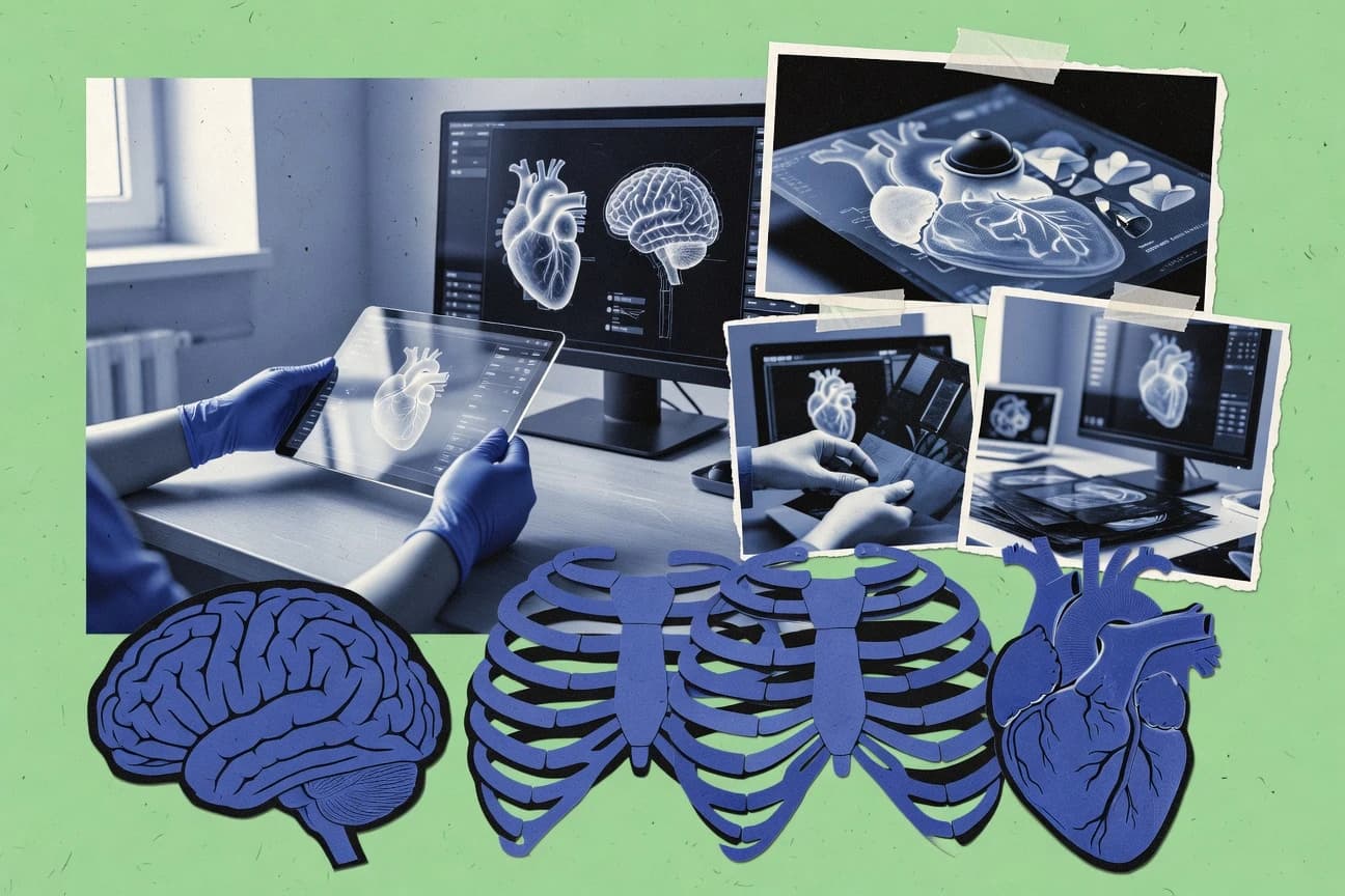
Top 10 3D Medical Software tools for imaging and analysis, ranked by features. Includes 3D Slicer and OsiriX MD for DICOM viewing.

How we ranked these tools
Core product claims cross-referenced against official documentation, changelogs, and independent technical reviews.
Analyzed video reviews and hundreds of written evaluations to capture real-world user experiences with each tool.
AI persona simulations modeled how different user types would experience each tool across common use cases and workflows.
Final rankings reviewed and approved by our editorial team with authority to override AI-generated scores based on domain expertise.
Score: Features 40% · Ease 30% · Value 30%
Gitnux may earn a commission through links on this page — this does not influence rankings. Editorial policy
Editor’s top 3 picks
Three quick recommendations before you dive into the full comparison below — each one leads on a different dimension.
3D Slicer
MRML node-based data model that unifies images, segmentations, and transforms across modules.
Built for fits when teams need extensible segmentation and registration workflows with scripted automation..
DICOM 3D Viewer (dcm4che ImageViewer)
Editor pick3D series-based volume rendering that keeps study and series metadata consistent with dcm4che indexing.
Built for fits when mid-size teams need controlled 3D viewing tied to dcm4che archive governance and automation..
OsiriX MD
Editor pickSchema-linked 3D derived outputs maintain traceability to DICOM studies and series.
Built for fits when mid-size clinical teams need controlled 3D review workflow automation and DICOM-consistent data lineage..
Related reading
Comparison Table
The comparison table benchmarks 3D imaging and analysis tools by integration depth, data model, automation and API surface, and admin and governance controls. It contrasts how each tool handles DICOM and derived objects, what schema or extensibility paths it exposes, and how teams manage RBAC, audit logs, and configuration at scale. Readers can use the table to assess tradeoffs in throughput, automation coverage, and provisioning workflows across desktop and server-based viewers and authoring platforms.
3D Slicer
open-sourceOpen-source medical image computing platform that supports 3D visualization, segmentation, registration, and 3D model-based workflows for radiology and research.
MRML node-based data model that unifies images, segmentations, and transforms across modules.
The core loop centers on creating and editing segmentation objects, registering images, and rendering results through a scene graph of MRML nodes. The extension architecture enables integration depth by adding new importers, algorithms, and user interface modules that operate on the same node data model. Automation is supported through scripting that can drive headless or semi-headless processing via the same MRML scene state used in the GUI.
A key tradeoff is that governance controls such as RBAC and audit logs are not native to the application, so deployments typically rely on external OS access controls and wrapper services. Teams use it when throughput depends on reusable scripted pipelines and when custom algorithms need to be packaged as modules that share the MRML data schema.
- +MRML scene model keeps volumes, segmentations, and transforms interoperable
- +Plugin modules add import, processing, and UI components on shared node APIs
- +Scripting can automate GUI workflows by driving MRML state
- +Segmentation and registration tools operate on consistent data structures
- –Enterprise-style RBAC and audit logs are not built into the core app
- –Automation typically depends on scripting and packaging, not a managed job API
- –Headless deployments require orchestration outside the application
Best for: Fits when teams need extensible segmentation and registration workflows with scripted automation.
More related reading
DICOM 3D Viewer (dcm4che ImageViewer)
DICOM stackDICOM-focused imaging capabilities that support viewing and handling of medical images for 3D inspection workflows.
3D series-based volume rendering that keeps study and series metadata consistent with dcm4che indexing.
Teams fit this viewer when DICOM ingestion and archive operations already run on dcm4che and the viewing layer must follow the same data model. The viewer works against DICOM concepts of patients, studies, series, and instances, so metadata stays consistent across the workflow. The integration depth is strongest when provisioning and configuration live alongside dcm4che services that handle storage and access.
A tradeoff appears when environments require a viewer that is independent of dcm4che server governance, because configuration and API integration patterns follow the same ecosystem. A common usage situation is a QA or radiology engineering workflow where series selection, metadata filtering, and repeatable rendering must stay coupled to archive indexing. Another situation is automated QA pipelines where rendering output needs to be reproducible for the same stored series.
- +Tight alignment with dcm4che data model for patient, study, series, and instance metadata
- +3D volume rendering based on archived DICOM series for repeatable visualization
- +Ecosystem integration improves workflow consistency from archive to viewer
- +Extensibility and configuration patterns fit governed enterprise deployments
- –Best fit depends on dcm4che ecosystem alignment for provisioning and access patterns
- –Automation requires building around the archive and indexing interfaces, not only the viewer UI
- –Advanced governance needs more surrounding infrastructure than a self-contained viewer
Best for: Fits when mid-size teams need controlled 3D viewing tied to dcm4che archive governance and automation.
OsiriX MD
clinical viewerDICOM imaging workstation that enables interactive 3D reconstruction and visualization for clinical review and analysis.
Schema-linked 3D derived outputs maintain traceability to DICOM studies and series.
OsiriX MD organizes imaging data by DICOM study and series and keeps derived outputs tied to the originating acquisition context. 3D work focuses on consistent volume interaction across sessions, which reduces rework when teams review the same study at different timepoints. The configuration layer supports repeatable review and export flows, which helps when throughput matters and the same steps must run across many cases.
The main tradeoff is that automation and API usage tend to require tighter alignment with OsiriX MD’s expected schema and object lifecycle. A common fit is a radiology workflow where ingestion, annotation, and structured export need to stay consistent across sites and where automation should create predictable derived artifacts.
- +Study and series data model keeps derived 3D outputs traceable to source DICOM
- +3D volume navigation supports repeatable review steps across multi-session workflows
- +Configuration-driven flows reduce variation in annotation and export steps
- +Automation surface enables external orchestration around a defined object lifecycle
- –API automation depends on matching OsiriX MD’s schema expectations for derived objects
- –Complex custom workflows may require deeper configuration than ad hoc scripting
Best for: Fits when mid-size clinical teams need controlled 3D review workflow automation and DICOM-consistent data lineage.
More related reading
RadiAnt DICOM Viewer
DICOM viewerDesktop DICOM viewer that provides fast 2D and 3D visualization workflows for radiology image review.
Real-time 3D volume rendering with interactive measurements on DICOM series volumes.
RadiAnt DICOM Viewer is a desktop DICOM viewer focused on fast 3D volume rendering and interactive inspection across large study sets. The tool’s data model centers on DICOM series, windowing, and derived volume reconstruction for consistent geometry during segmentation and measurements.
Integration depth depends on how organizations externalize ingestion and labeling, because RadiAnt’s automation surface is primarily task scripting around local viewing workflows. Admin and governance controls are limited to local workstation usage patterns rather than centralized RBAC, audit log, or provisioning controls.
- +Interactive 3D volume rendering tuned for local workstation throughput
- +Consistent DICOM series handling supports repeatable measurements and review
- +Extensible workflow via scripting and configuration of viewer behavior
- –Automation and API surface are narrower than server-based DICOM platforms
- –Centralized RBAC, audit log, and provisioning controls are not the focus
- –Governance for multi-site workflows relies on external process controls
Best for: Fits when imaging teams need high-throughput local 3D viewing and measurement without centralized governance.
Horos
desktop viewerFree macOS DICOM viewer that supports multi-planar and 3D rendering workflows for medical image interpretation.
Horos add-on architecture for extending 3D visualization, segmentation tools, and annotation workflows.
Horos provides an open-source DICOM viewer that supports 3D medical image rendering and advanced visualization workflows. Its extension model lets installations add tooling without changing the base viewer, which affects configuration and extensibility.
Integration depth centers on DICOM connectivity and exported artifacts such as images, segmentations, and measurements for downstream analysis. Automation and API surface depend on how extensions are built for provisioning, schema alignment, and RBAC boundaries in the surrounding clinical environment.
- +DICOM-focused data model with consistent studies, series, and instances
- +Extensibility via add-ons that change visualization and annotation behavior
- +3D rendering supports segmentation-driven views and measurements
- +Exportable outputs enable integration with PACS workflows and pipelines
- –API surface is mainly extension-based, not built for external automation
- –RBAC and audit log capabilities rely on deployment around the viewer
- –Automation throughput depends on workstation orchestration, not server queues
- –Schema alignment for derived data needs custom conventions by integrators
Best for: Fits when teams need a configurable DICOM viewer with 3D rendering and extension-based automation.
More related reading
ParaView
visualizationVisualization application that supports scientific 3D rendering and can be used for medical volume visualization outputs and custom pipelines.
ParaView Python scripting with ParaView server-client batch execution for automated rendering and pipeline processing.
ParaView fits teams running medical visualization pipelines that need scripted reproducibility and extensibility through plugins and Python automation. The data model centers on VTK objects, with readers and filters that map to a clear processing graph for consistent 3D rendering outputs.
Integration depth comes from Python scripting, ParaView server-client execution, and a documented extension mechanism for custom sources, filters, and writers. Automation and API coverage are strongest for workflow orchestration via Python, while admin governance depends largely on how the deployment wraps access control and shared resources.
- +VTK-based pipeline graph enables reproducible 3D processing runs
- +Python automation covers headless rendering, batch filters, and pipeline setup
- +Plugin extensibility adds custom readers, filters, and output writers
- +Client-server mode supports remote compute for large medical volumes
- –Built-in governance such as RBAC and audit logging is not a native focus
- –Automation relies heavily on scripting, which increases maintenance overhead
- –Cross-system integration needs custom glue for authentication and job orchestration
- –Large datasets can stress memory and throughput without careful pipeline design
Best for: Fits when engineering teams need scriptable 3D medical visualization with extensibility and pipeline reproducibility.
NVIDIA Clara Imaging
GPU imagingMedical imaging visualization and developer tools designed for GPU-accelerated DICOM ingestion and 3D visualization in imaging pipelines.
Clara container and workflow composition enables configurable, repeatable imaging pipeline assembly.
NVIDIA Clara Imaging differentiates through its containerized, Kubernetes-ready deployment model for imaging data workflows and inference pipelines. The core capabilities center on a curated set of imaging applications plus an extensible integration approach via Clara containers, SDK components, and documented interfaces for assembling 3D medical workflows.
The data model and schema design follow imaging-centric conventions that support consistent preprocessing, inference input shaping, and output packaging for downstream consumption. Integration depth is driven by orchestration patterns, configuration injection, and an automation surface intended for repeatable provisioning and pipeline throughput across environments.
- +Container-first architecture fits Kubernetes orchestration for imaging pipelines
- +Extensible integration via Clara components for assembling multi-stage 3D workflows
- +Configuration-driven deployment supports repeatable environment provisioning
- +Workflow outputs are packaged for downstream stages and system integration
- –Integration effort increases when replacing default Clara pipeline components
- –RBAC and governance controls depend on external Kubernetes and platform layers
- –Data schema alignment can be nontrivial when mixing imaging sources
- –Debugging spans containers and orchestrator logs across multiple services
Best for: Fits when teams need containerized 3D medical workflows with automation-first deployment and integration control.
More related reading
Materialise Mimics
medical modelingMedical image processing software that converts 2D slices into 3D masks and models for analysis, design, and manufacturing workflows.
Advanced segmentation tools with measurement and model export for consistent, reusable 3D outputs.
Materialise Mimics centers on a clinical imaging to 3D segmentation workflow tied to an explicit data model for medical surfaces, volumes, and measurement outputs. Integration depth is largely driven through exportable artifacts, scripting and automation hooks, and interoperability with Materialise tooling for downstream planning and review.
The automation surface supports repeatable preprocessing, segmentation, and reporting steps, with extensibility via APIs and connectable pipelines in common imaging environments. Data governance relies on project-level controls, user roles, and traceable operations to support auditability across teams handling regulated artifacts.
- +Segmentation-to-mesh pipeline outputs consistent surface and volume artifacts for downstream planning
- +Strong interoperability via import export formats used in clinical imaging workflows
- +Automation support for repeatable segmentation and measurement workflows
- +Extensibility through scripting and integration points into connected processing pipelines
- –Integration is more artifact-oriented than full schema synchronization across systems
- –Automation coverage depends on available scripting hooks for each workflow stage
- –Admin governance granularity may not match enterprise IAM needs without additional layers
- –Throughput gains from batch processing depend on external orchestration around Mimics
Best for: Fits when clinical teams need controlled, automatable segmentation artifacts for downstream medical workflows.
Conclusion
After evaluating 8 healthcare medicine, 3D Slicer stands out as our overall top pick — it scored highest across our combined criteria of features, ease of use, and value, which is why it sits at #1 in the rankings above.
Use the comparison table and detailed reviews above to validate the fit against your own requirements before committing to a tool.
How to Choose the Right 3D Medical Software
This buyer's guide explains how to choose 3D Medical Software for 3D visualization, segmentation, registration, and measurement workflows. It covers open platforms like 3D Slicer, DICOM-first viewers like RadiAnt DICOM Viewer and DICOM 3D Viewer from dcm4che ImageViewer, and production-style pipeline tools like NVIDIA Clara Imaging and Materialise Mimics. It also clarifies where visualization-only tools like ParaView fit versus clinical workstation tools like OsiriX MD and Horos.
What Is 3D Medical Software?
3D Medical Software turns medical imaging data such as CT and MRI stacks into interactive 3D views, multiplanar reconstructions, and measurement outputs. These tools help users inspect anatomy, quantify structures, and convert image slices into segmentation masks and watertight 3D models. In practice, 3D Slicer provides a full clinical-grade 3D visualization and segmentation workflow with modules like Segment Editor. For DICOM-focused viewing, RadiAnt DICOM Viewer and DICOM 3D Viewer from dcm4che ImageViewer combine interactive 3D volume rendering with multiplanar reconstruction for rapid radiology-style review.
Key Features to Look For
The right feature set depends on whether the workflow centers on inspection, quantitative measurement, segmentation and reconstruction, or GPU-driven production pipelines.
Interactive 3D volume rendering paired with multiplanar reconstruction
Look for tools that show true 3D rendering and allow multiplanar reconstruction without switching applications. DICOM 3D Viewer from dcm4che ImageViewer combines interactive multiplanar reconstruction with 3D volume rendering for DICOM image stack review, and RadiAnt DICOM Viewer delivers realtime multiplanar reconstruction with instant responsiveness.
Segmentation workflows with multiple segmentation effects and live 3D preview
Choose segmentation that supports multiple approaches and provides immediate 3D feedback so corrections happen during editing. 3D Slicer stands out with Segment Editor that includes multiple segmentation effects and live 3D preview controls. Horos also pairs segmentation tools with multiplanar reformatting for accurate 3D anatomical review.
Robust registration and transform management for multi-modal alignment
Select registration features when overlaying images from different time points, scanners, or modalities matters. 3D Slicer includes robust registration tools for multi-modal alignment and transform management. NVIDIA Clara Imaging is designed for GPU-accelerated segmentation and registration pipeline building via Clara Imaging SDK.
Quantitative measurement and annotation for clinical-style review
For radiology work, measurement and annotation tools must support fast, consistent analysis during inspection. RadiAnt DICOM Viewer provides measurement and annotation tools tied to fast 3D MPR navigation. OsiriX MD supports measurement and annotation workflows alongside DICOM-aware 3D volume rendering and multiplanar reconstruction.
Reproducible visualization pipelines with scripted filter graphs
Researchers who need repeatable outputs should prioritize pipeline-driven visualization. ParaView provides a filter pipeline for structured volume rendering and uses Python scripting to automate visualization and analysis steps. ParaView can also use parallel and distributed rendering options to handle large volumetric datasets.
Watertight image-to-model conversion with mask-based editing and geometry export
Teams that need 3D models for analysis, design, or manufacturing require robust mask-based segmentation and geometry editing. Materialise Mimics focuses on converting CT and MRI slices into analysis-ready 3D masks and models with interactive segmentation and precise measurement tools. It also supports mesh and geometry editing for downstream CAD-style edits and medical device design handoffs.
How to Choose the Right 3D Medical Software
A practical decision starts by matching the workflow goal to the tool strengths in 3D viewing, segmentation and reconstruction, or production-grade processing.
Start with the intended workflow output
If the goal is radiology-style inspection with fast 3D and multiplanar navigation, RadiAnt DICOM Viewer and OsiriX MD fit because they deliver realtime 3D MPR and measurement-ready review within the viewer. If the goal is clinical-grade segmentation and quantitative analysis inside a configurable workspace, 3D Slicer is a strong match due to Segment Editor with multiple segmentation effects and live 3D preview.
Verify DICOM-focused controls for the datasets used in day-to-day work
DICOM-first teams should check that study navigation and rendering are built around DICOM image stacks. DICOM 3D Viewer from dcm4che ImageViewer integrates tightly with the dcm4che ecosystem and supports metadata-driven workflow navigation with multiplanar reconstruction. RadiAnt DICOM Viewer is built for local file handling workflows with fast interactive 3D rendering.
Evaluate segmentation depth versus model-generation requirements
Segmentation for measurement often prioritizes accurate labeling and quick corrections during editing. 3D Slicer supports segmentation, labeling, and interactive measurement with a modular architecture. If the output must be a watertight model for downstream geometry, Materialise Mimics adds mask-based editing and geometry preparation for CAD-style handoffs.
Assess whether reproducibility and scripting are mandatory
Teams that need repeatable rendering and structured processing should select ParaView because it uses a filter pipeline and Python scripting for automated visualization steps. For teams that need end-to-end pipeline building with GPU acceleration, NVIDIA Clara Imaging focuses on Clara Imaging SDK GPU-optimized imaging libraries for segmentation and registration workflows.
Match deployment realities to the tool’s operating model
For mixed OS clinical environments, pick tools that align with the team’s platform constraints because OsiriX MD is centered on a Mac-based workflow. If the goal is research extensibility, 3D Slicer and Horos provide plugin-driven ecosystems that extend capabilities without rebuilding the core app. If the goal is engineering integration, NVIDIA Clara Imaging expects engineering effort to integrate imaging modules into existing systems.
Who Needs 3D Medical Software?
Different 3D Medical Software tools target distinct roles across clinical review, research pipelines, and production image-to-model workflows.
Clinical research teams building segmentation, registration, and analysis workflows
3D Slicer fits because it combines registration tools with a Segment Editor that supports multiple effects and live 3D preview controls. ParaView can complement 3D Slicer when research teams need reproducible filter-pipeline rendering and Python automation.
Radiology teams that need fast DICOM 3D viewing with measurement during review
RadiAnt DICOM Viewer matches this need because it delivers realtime multiplanar reconstruction with interactive 3D navigation and instant responsiveness. OsiriX MD also supports DICOM-aware 3D volume rendering plus multiplanar reconstruction and measurement workflows on macOS.
Mac-based radiology and research users focused on DICOM visualization and analysis
Horos is designed as a free macOS DICOM viewer that supports multiplanar reformatting and 3D volume rendering. It also includes segmentation tools combined with multiplanar reformatting to support accurate 3D anatomical review.
Teams converting CT and MRI into analysis-ready or manufacturing-ready 3D models
Materialise Mimics is built for interactive segmentation and mask-based editing that converts CT and MRI into accurate 3D masks and models. It supports robust measurement for distances and volumes and provides mesh and geometry editing for downstream CAD-style edits and device design handoffs.
Common Mistakes to Avoid
Misalignment between workflow goals and tool strengths leads to slow setups, incomplete outputs, or extra manual steps across these 3D Medical Software options.
Choosing a segmentation tool without validating workflow complexity
3D Slicer and Horos provide advanced segmentation capabilities, but both can create a steep learning curve for newcomers and require careful setup for segmentation workflows. ParaView also introduces complexity via a filter graph that can slow first-time setup when segmentation and visualization are not yet standardized.
Expecting a DICOM viewer to replace a full imaging workstation
DICOM 3D Viewer from dcm4che ImageViewer focuses on viewing and annotation-style analysis and does not provide a complete PACS-grade imaging feature set. RadiAnt DICOM Viewer also emphasizes local review workflows and does not deliver collaboration and reporting depth comparable to PACS-grade platforms.
Selecting a visualization pipeline tool for interactive clinical editing
ParaView is powerful for structured volume rendering pipelines and Python-driven repeatability, but it is not positioned as a turn-key clinical workstation for interactive segmentation and correction. 3D Slicer and Materialise Mimics better match interactive segmentation and model preparation needs for clinical and engineering workflows.
Assuming GPU-accelerated libraries remove integration and validation work
NVIDIA Clara Imaging provides Clara Imaging SDK GPU-optimized imaging libraries for segmentation and registration workflows, but it still requires engineering effort to integrate modules into existing systems. This can increase validation overhead for regulated use cases, so integration planning must start early.
How We Selected and Ranked These Tools
we evaluated every tool on three sub-dimensions that directly map to buying priorities: features with a weight of 0.4, ease of use with a weight of 0.3, and value with a weight of 0.3. the overall rating is the weighted average of those three sub-dimensions computed as overall = 0.40 × features + 0.30 × ease of use + 0.30 × value. 3D Slicer separated itself because it scored strongly on the features dimension by combining clinical-grade 3D visualization with segmentation through Segment Editor and live 3D preview controls. this feature breadth supported more end-to-end workflows than DICOM-only viewing tools and provided deeper extensibility than pipeline-only visualization tools.
Frequently Asked Questions About 3D Medical Software
Which tool fits most imaging-to-segmentation workflows that need repeatable scripting?
How do 3D Slicer and Horos differ when teams need to extend 3D visualization and labeling?
What integration path is strongest when an organization already runs a dcm4che archive?
Which platform is better for building automated 3D rendering pipelines with custom processing stages?
How does OsiriX MD maintain traceability compared with RadiAnt DICOM Viewer during review and reporting?
Which tool is more suitable for converting segmentation and measurements into artifacts for downstream medical planning workflows?
What admin control expectations should teams set for 3D Slicer versus dcm4che ImageViewer or OsiriX MD?
When teams hit data migration issues, what does the data model imply for moving studies and derived objects?
Which toolchain suits organizations that need controlled configuration to prevent inconsistent 3D viewing outputs?
Tools reviewed
Primary sources checked during evaluation.
Referenced in the comparison table and product reviews above.
Keep exploring
Comparing two specific tools?
Software Alternatives
See head-to-head software comparisons with feature breakdowns, pricing, and our recommendation for each use case.
Explore software alternatives→In this category
Healthcare Medicine alternatives
See side-by-side comparisons of healthcare medicine tools and pick the right one for your stack.
Compare healthcare medicine tools→FOR SOFTWARE VENDORS
Not on this list? Let’s fix that.
Our best-of pages are how many teams discover and compare tools in this space. If you think your product belongs in this lineup, we’d like to hear from you—we’ll walk you through fit and what an editorial entry looks like.
Apply for a ListingWHAT THIS INCLUDES
Where buyers compare
Readers come to these pages to shortlist software—your product shows up in that moment, not in a random sidebar.
Editorial write-up
We describe your product in our own words and check the facts before anything goes live.
On-page brand presence
You appear in the roundup the same way as other tools we cover: name, positioning, and a clear next step for readers who want to learn more.
Kept up to date
We refresh lists on a regular rhythm so the category page stays useful as products and pricing change.